

 PDF
PDF ePub
ePub Citation
 Print
Print


INTRODUCTION
Benign anastomotic stricture after upper gastrointestinal (UGI) surgery can be classified as early (within 3 months after operation) and late (more than 3 months after operation) (1). Early anastomotic strictures are usually secondary to edema, whereas late strictures are related with fibrotic scarring (12). Balloon dilatation is regarded as a safe and effective initial treatment for benign strictures (34). In cases of benign stricture refractory to balloon dilation or immediate recoil after balloon dilation, retrievable metallic stent placement can be used as a treatment option (5).
A covered retrievable stent can be removed several weeks after placement. However, it has a high risk of stent migration (6). Stent migration rate in benign disease is higher than that in malignant disease. According to published data, the rate of stent migration is 25 to 60% for benign disease and 7 to 35% for malignant disease (789). Proximally migrated stent can be easily removed under endoscopy or fluoroscopy. In contrast, it is difficult or impossible to remove a distally migrated stent beyond the Treitz ligament under endoscopy and/or fluoroscopy. Most distally migrated stents pass through the rectum without any complications. However, distally migrated stents can cause intestinal obstruction or perforation (1011121314).
To prevent distal migration of the stent and possible complications such as intestinal perforation, the concept of a fixation string secured to the patient's ear or nose has been introduced (1516). The purpose of this study was to evaluate the efficacy of retrievable metallic stent with a fixation string for benign stricture after UGI surgery.
MATERIALS AND METHODS
Patients
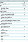
From June 2009 to May 2015, we placed 56 self-expandable retrievable stents with fixation string in 42 patients (mean age, 60.8 years; age range, 34–81 years) with early benign stricture after UGI surgery. Inclusion criteria were: 1) patients underwent UGI surgery within 3 months, 2) benign stricture demonstrated radiologic study, 3) clinical symptom of indigestion, 4) treatment with retrievable stent. Exclusion criteria were: 1) patients underwent esophageal surgery, 2) stricture was caused by malignant disease.
There were 25 males (mean age, 60.7 years; age range, 35–76 years) and 17 females (mean age, 60.8 years; age range, 34–81 years). The cause of surgery was benign disease in 10 patients and malignant disease in 32 patients. Twelve patients who had received pylorus preserving gastrectomy were reported in a previous paper focusing on the management of post-operative pyloric spasms (17).
Upper gastrointestinal study was performed at 3 to 60 days (mean, 17.2 days) after operation. Benign stricture was defined as a narrowing segment maintaining a normal mucosal fold without a shouldering edge. Out of 42 patients, 35 had undergone balloon dilatation (balloon catheter of 18–25 mm in diameter) prior to stent placement. Due to immediate recoil or no improvement of diet, stent placement was required. Seven patients underwent primary stent placement due to severe long segmental stenosis on UGI study. The interval from surgery to stent placement was 5 to 65 days (mean, 19 days; median, 20 days). The demographic characteristics of these patients are summarized in Table 1.
Stent Construction
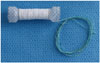
The stent used in this study was a covered retrievable Niti-S stent (Taewoong Medical, Gimpo, Korea). It was composed of a single thread of 0.1778 mm nitinol wire shaped with wide-diameter proximal and distal ends in a dumbbell configuration. The stent was fully covered with silicone and polytetrafluoroethylene. A polyester string of 150 cm in length was wrapped around the proximal end of the stent body (Fig. 1). The string was inside the introducer sheath. The end of the string exited through the side hole of the handle.
Stent Placement Techniques
Topical anesthesia of the pharynx was performed routinely using aerosol spray (Xylocaine; Astrazeneca Korea, Seoul, Korea) before stent placement procedure. Under fluoroscopic guidance, a 5F catheter and 0.035" hydrophilic guidewire (Terumo, Tokyo, Japan) were inserted through the mouth and advanced across the estimated stricture site. A small amount of water-soluble contrast medium was then injected while withdrawing the catheter to outline the stricture. After that, a 0.035" exchange stiff guidewire (Terumo) or extra-stiff guidewire (Lunderquist, Cook medical, Bloomington, IN, USA) was inserted and advanced across the stricture site. The stent delivery system was advanced over the guidewire. The retrievable stent of 8–15 cm in length and 20 mm in diameter was released over the stricture. After the stent was released, the string was passed through the mouth using the delivery system. A Nelaton tube (Qingdao Sewon Medical, Qingdao, China) was then inserted into the nostril and pulled out of the mouth using forceps. The string was tied to the Nelaton tube. By pulling the Nelaton tube, the string would come out of the nostril. After the Nelaton tube was cut off, the end of the string was tied to a 0.014-inch micro-guidewire (Transend; Boston Scientific, Natick, MA, USA). The micro-guidewire and the string were then passed through the Nelaton tube to reduce pain in the nostril caused by the string. After the micro-guidewire was cut off, the end of the string was wound around the gauze and anchored around the ear using a tape (Fig. 2).
To confirm the position of the stent, all patients underwent daily plain radiograph. We also checked whether they felt pain around the ear and nostril as the string was pulled down due to the stent with distal migration.
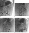
Removal of the stent was performed two weeks after the stent placement or when stent-related complications such as stent migration occurred. The string in the oral pharynx was grabbed with forceps and pulled out of the mouth. The end of the string was tied to 0.014-inch micro-guidewire (Transend). A 7-Fr guiding catheter (Envoy; Cordis, Miami Lakes, FL, USA) was then advanced over the micro-guidewire and the string to reach the proximal end of the stent. When the string was pulled, the proximal end of the stent was collapsed and the stent was retrieved (Supplementary Movie 1 in the online-only Data Supplement). Repositioning of the distally migrated stent was performed using a fashion similar to the removal technique (Fig. 3).
Analysis
Technical success was defined as successful deployment of the stent across the stricture with confirmation of patency using fluoroscopy. To assess clinical success, food intake capacities before and after stent placement were determined. Food intake capacity was categorized as non per os, sips of water, soft fluid diet, soft blend diet (SBD), and normal regular diet (NRD). Clinical success was defined as achieving NRD.
Migration was categorized into the following four groups: complete proximal migration, partial proximal migration, complete distal migration, and partial distal migration. Complete migration was defined as migration of the stent completely out of the stricture, whereas partial migration was defined as stent remaining partially within the stricture.
Independent t test was used to compare the stent indwelling time between the group that achieved clinical success with additional procedure after the initial stent removal was performed and the group that achieved clinical success without additional procedure after the initial stent removal. Statistical analysis was carried out using MedCalc for Windows, version 15.8 (MedCalc Software, Mariakerke, Belgium). Statistical significance was considered when p value was less than 0.05.
RESULTS
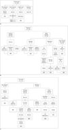
Of the 42 initial stents, 15 (35.7%) showed no migration. Malfunction was observed in 3 (7.1%) stents due to food impaction in two patients and stent kinking in one patient. Stent migration occurred in 24 (57.1%) initial stents (Fig. 4A).
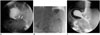
All 15 stents without migration were electively removed under endoscopy or fluoroscopy guidance. Indwelling periods for these stents were 14 to 21 days in 14 patients and 7 days in one patient for whom the operator removed the stent without specific reason. All these 15 patients achieved NRD after stent removal (Fig. 5).
Two food-impacted stents were removed at 4 days and 7 days after placement, respectively. Without any additional procedure, one patient achieved NRD and the other patient achieved SBD. Stent malfunction due to stent kinking occurred in one patient. She had undergone Billroth II subtotal gastrectomy. The stent was placed at the anastomosis. However, the stent was collapsed immediately after placement. It did not improve on serial radiographs. The stent was removed at 7 days after placement. Food intake was not improved. Surgical management was carried out and an adhesion band around the anastomosis was revealed. This patient was able to achieve NRD after adhesiolysis.
Of the 24 migrated initial stents, 15 migrated proximally (partial, 3; complete, 12) and 9 migrated distally (partial, 2; complete, 7) (Fig. 4B). The period between stent placement and the occurrence of migration ranged from 1 to 14 days (mean, 3.6 days). All 15 proximally migrated stents were successfully removed. After stent removal, three patients achieved NRD without additional procedure, three patients underwent surgery (adhesiolysis in two patients and gastrojejunostomy in one patient), and the remaining nine patients underwent second stent placement.
Of the 9 distally migrated stents, one was discharged through the rectum at 14 days after placement without any complication and three were successfully removed. Repositioning of migrated stents was attempted for the remaining five stents. Out of the three patients with successful stent removal, one achieved NRD without additional procedure, one underwent second stent placement, and one achieved NRD after one session of balloon dilatation. Repositioning of the stent was successful in four of the five patients. In one patient, repositioning of the stent failed. Therefore, the stent was removed immediately. The patient underwent a second stent placement and two sessions of balloon dilatation. However, food intake did not improve. He achieved NRD after gastrojejunostomy.
All four successfully repositioned stents migrated distally again. Of these, two were successfully removed. The two patients achieved NRD without any additional procedure. For the remaining two patients, the removal of migrated stents failed because the operators lost the strings during retrieval. One patient achieved NRD without additional treatment and the stent passed through the rectum without complications. The other patient underwent surgical adhesiolysis due to persistent obstructive symptoms. The migrated stent was removed during surgery.
Eleven patients (nine patients with proximally migrated initial stents and two patients with distally migrated initial stents) received repeated stent placement (a total of 14 stents) (Fig. 4C). Of the 11 second-stents, 3 did not migrate. The three patients achieved NRD after successful elective stent removal. Eight of the 11 second-stents migrated (six proximally and two distally). All proximally migrated second-stents were removed successfully. After the removal, three patients achieved NRD, one achieved NRD after one session of balloon dilatation, and two underwent a third stent placement. Of the two distally migrated second-stents, one stent was repositioned but migrated distally again. It was successfully removed. The patient achieved NRD without any additional procedure. The other distally migrated second-stent was removed and the patient achieved NRD after two sessions of balloon dilatation and gastrojejunostomy. Of the two third-stents, one migrated proximally and the other migrated distally. These stents were successfully removed. One patient achieved NRD after stent removal. The other patient underwent a fourth stent placement. In this patient, the fourth stent was removed electively without migration. He achieved NRD.
In summary, 56 stents were placed in 42 patients. The technical success rate was 100%. The clinical success rate after the first stent placement was 57.1% (24/42). The clinical success rate after repeated stent placement and/or balloon dilation was 83.3% (35/42). Six (14.3%) patients achieved NRD after surgical correction. Stent migration occurred in 60.7% (34/56) of patients (Table 2). Stent removal was successful in 94.6% (53/56) of patients. Distal migration occurred in 12 stents. Among these, 10 (83.3%) stents were successfully removed whereas two stents could not be removed.
Among 35 patients who achieved NRD after stent placement and/or balloon dilation, the indwelling time of the initial stent was longer (p < 0.0001) in patients with clinical success after the first stent placement than that in patients with clinical success after repeated stent placement and/or balloon dilation (Table 3).
DISCUSSION
Anastomotic stricture occurs in 3 to 13% of patients after gastric surgery and in 18–50% of patients after esophagectomy (3). Early anastomotic strictures (within 3 months after the operation) are usually secondary to postoperative edema, while late strictures (more than 3 months after the operation) are related to fibrotic scarring (12). Anastomotic stricture can be treated with fluoroscopy or endoscopy-guided balloon dilatation, covered stent placement, or surgical revision (131819). Balloon dilatation has been regarded as an initial treatment for benign anastomotic stricture. According to previously reported data, fluoroscopy-guided balloon dilatation is effective in 95% of anastomotic stricture after Ivor-Lewis esophagectomy and 94% of total gastrectomy with Billroth I or II (1920). However, fibrotic stricture or tortuous and angulated stricture such as those at the gastrojejunostomy site may be refractory to balloon dilatation (120). In such refractory cases, temporary placement of a covered metal stent can be an effective option (6212223). The covering of covered stents limits tissue ingrowth and allows for easy removal (6). However, it can increase the rate of stent migration compared to uncovered stents (6). In addition, the stent migration rate is higher in benign stricture than in malignant stricture (25–60% vs. 7–35%) (789). A few studies have reported that anastomotic stricture is more commonly associated with stent migration than other benign causes (16–30%), although there is no statistical significance (724). Stents for gastroesophageal junction and pylorus stricture can also increase the likelihood of migration (2526).
If the migrated stent remains in the stomach, endoscopic removal of the stent is possible. However, removal of a more distally migrated stent is difficult or impossible. Most distally migrated stents can spontaneously pass through the rectum or remain in the body without any complications (7). In a few cases, however, distally migrated stents have caused intestinal perforation or obstruction leading to surgery (1011121314). Retrieval hooks have been used to remove covered stents with drawstrings. However, it is also impossible to remove stents that have migrated too distally using a retrieval hook (727). Thus, to prevent migration of covered stents into the distal jejunum and facilitate the removal of migrated stents, covered stents with a long string anchoring to the ear of patient has been developed. This study determined the efficacy of using a fixation string to remove or reposition the distally migrated stent. A total of 10 out of 12 distally migrated stents were successfully removed using a fixation string and angiocatheter.
The migration rate of our stents was 60.7%, which was slightly higher than those reported in other studies (5728). Such difference in migration rate might be due to different patient characteristics between this study and other studies. Patients in other studies had benign conditions such as radiation fibrosis, corrosive stricture, and achalasia (5728), whereas the present study included only early anastomotic stricture after UGI surgery. The stenotic lesion of radiation fibrosis and corrosive stricture is relatively tight and long in length. However, anastomotic stenosis commonly represents a short segmental discrete lesion, which might result in more common stent migration at the anastomotic site. The straight course of anastomosis also can influence stent migration. The gastrojejunostomy site is more angulated than the gastroduodenostomy site. Therefore, migration is more common in gastroduodenostomy than that in gastrojejunostomy. (20)
The advantage of using a retrievable stent with a fixation string is that the distally migrated stent can be removed or repositioned using the string and angiocatheter. This can decrease the risk of bowel obstruction or perforation caused by the distally migrated stent. Repositioning of distally migrated stents can increase the indwelling time of stents and obviate the need for additional stents. However, patients might have pain in their nostrils or ears due to the string. An inexperienced operator may lose the string during stent removal or repositioning, resulting in failure of stent removal.
The optimal indwelling time of the retrievable stent has not been established. Song et al. (29) have reported that stent migration after 2 months does not form new strictures. The optimal duration for stent placement would be 4–8 weeks (29). We removed the stent with a string electively two weeks after the placement if the stent showed no migration. No patient suffered from recurrence of the stricture after stent removal. Song et al. (29) have mainly dealt with corrosive stricture of the esophagus. Because corrosive stricture is a tight fibrotic stricture, sufficient indwelling time of the stent is required. In contrast, early anastomotic stricture is an edematous lesion. Therefore, shorter indwelling time of the stent may be sufficient. We believe that 2 weeks of stent indwelling time is enough for early anastomotic stricture.
This study has several limitations. First, this study was retrospectively conducted in a single center with a small number of patients. Second, retrievable stent placement after esophageal surgery was not included. Experiences are required to obtain clinical result of retrievable stent placement regarding other anastomotic sites. Third, because this is a retrospective study, we were unable to evaluate the pain scores of patients' nostrils and/or ears. Fourth, in this study, 35 patients received balloon dilatation before stent placement whereas 7 patients received primary stenting due to severe stenosis. Patients underwent various kinds of surgeries with diverse clinical course. Thus, neither standard treatment algorithm nor significant factors affecting clinical outcome could be retrieved from this study. Fifth, patients were usually discharged when they could tolerate SBD after stent removal. Thus, this retrospective study was not able to determine the exact time when the patient achieved NRD after the stent removal.
In conclusion, placement of a retrievable covered stent with a fixation string is a feasible option for managing early benign anastomotic stricture after UGI surgery. Complications from distally migrated stents might be reduced by using a retrievable stent with a fixation string.


 XML Download