

 PDF
PDF ePub
ePub Citation
 Print
Print


INTRODUCTION
Radiofrequency (RF) ablation is an important therapeutic tool for the treatment of primary and secondary liver malignancies (1234). Complete necrosis is achieved in over 90% of cases of small hepatocellular carcinomas ≤ 3 cm in size (45). Results of treatment of very-early and early hepatocellular carcinomas is reportedly comparable to surgery (67). Various complications related to RF ablation of liver tumor have been reported, but the procedure is considered as relatively safe with overall complication rate of 7.2–12% (891011). When the tumor is located at high-risk locations in close proximity to the large vessel, bile duct, extra-hepatic organ or the liver capsule, the incidence of complication may increase. Subsequently, the procedure needs to be performed cautiously or in conjunction with additional special techniques (121314).
Radiofrequency ablation has the disadvantage that obtaining adequate length of ablation margin in all directions may be difficult for index tumor located at the superficial subcapsular region of the liver. In addition, reported incidence of electrode tract seeding is reportedly as high as 12.5% in patients with subcapsular tumors (15). Although subsequent reports suggest much lower incidence of seeding (891011161718192021), RF ablation is generally not a preferred procedure for subcapsular tumors (141516). Subsequently, it was recommended to avoid direct insertion of the electrode into the tumor without traversing normal parenchyma (22). A technique of ablation of subcapsular tumor without direct insertion of the antenna during microwave ablation has also been reported (23).
To obtain sufficient length of ablation margin in all directions and to traverse non-tumorous liver parenchyma during RF ablation, we modified the cool-tip electrode so that it is angled at the junction of insulated and exposed segment. The angled cool-tip electrode was used for RF ablation of small subcapsular tumors abutting the abdominal wall. In this study, we reported the feasibility and results of using the angled cool-tip electrode for RF ablation of superficially located small subcapsular tumors in liver abutting the abdominal wall.
MATERIALS AND METHODS
Patient Population
This retrospective study was approved by the Institutional Review Board, and written informed consent for the procedure was obtained from all patients. Requirement of informed consent prior to inclusion was waived due to the retrospective nature of the study. All procedures were performed in accordance with the guidelines of the institution.
From March 2013 to June 2015, among 342 RF ablations for primary and secondary liver malignancies, angled cool-tip electrode was used for RF ablation of 15 superficial subcapsular tumors of 3 cm or smaller in the longest dimension in 15 patients, and was retrospectively evaluated. The patients were 12 men and 3 women with ages ranging from 37 to 69 years (mean, 52.60 ± 8.78 years).
The index tumors were hepatocellular carcinoma (n = 13), metastasis from rectosigmoid cancer (n = 1) and metastasis from cholangiocarcinoma (n = 1). All tumors were newly developed lesions, superficially located, abutting the liver capsule in the abdominal wall side, diagnosed by clinical findings and dynamic CT and/or MRI during follow-up, with the diameter in the longest dimension measuring 10–26 mm (mean, 15.68 ± 5.29 mm). Of the 13 patients with hepatocellular carcinoma, all patients had underlying liver cirrhosis, related to B-viral hepatitis in 10, C-viral hepatitis in 1 and alcoholic cirrhosis in 2 patients. The hepatocellular carcinomas were diagnosed by typical image findings of enhancement on arterial phase with washout on portal or delayed phase (n = 13), and combined increased tumor markers (alpha-fetoprotein [α-FP] > 400, α-FP-L3% > 10, or proteins induced by vitamin K absence or antagonist-II [PIVKA-II] > 40) (n = 9) (2425). The metastasis from cholangiocellular carcinoma was diagnosed by increased size of the tumor on 2 month follow-up liver MRI with increased carcinoembryonic antigen (CEA) level, and the metastasis from rectosigmoid cancer was diagnosed by CT and MRI findings consistent with metastasis and increasing CEA level on follow-up. Positron emission tomography was not performed in patients with metastasis. In all patients, histological diagnosis of the tumor was not performed.
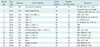
The angled cool-tip electrode was used for RF ablation in any primary or secondary liver tumors that was single in number, with diameter of 3 cm or smaller in the longest dimension in both the axial and the coronal images on liver dynamic CT and/or MRI, and located at superficial supcapsular region of the liver abutting the abdominal wall. Exclusion criteria included any tumor abutting critical organ such as bowel or gall bladder, uncooperative patient, and uncontrolled bleeding tendency. Characteristics of the patients and the tumors were summarized in the Table 1.
RF Ablation Technique
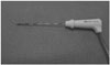
In all cases, the 17 G cool-tip electrode of 15 cm length with 3 cm exposed tip (Covidien, Mansfield, MA, USA) was used to prepare the angled electrode. In all cases, the angle was made toward the opposite direction where the tubes and the electrical wire was attached. Using a sterile needle holder, the electrode was angled manually to an angle of 25–35° by adding 4–5 small bends within 5–6 mm length of the electrode, centered at the junction of insulated and exposed segment of the electrode (Fig. 1).
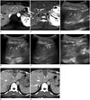
Pre-RF ablation ultrasonography evaluation was performed to determine the skin access site. An imaginary target point within the normal liver parenchyma at the depth of the epicenter of the index tumor and at about 1 cm apart from the tumor margin was selected. From this target point, an imaginary line with about 30° angle from the skin of the patient toward the farther side from the index tumor was drawn. The point where the imaginary line crossed the patient's skin was determined as the skin access site of the angled cool-tip electrode. After sterile preparation of the patient's skin, the pre-determined access site was anesthetized using 1% lidocaine hydrochloride. By holding the proximal, non-angled segment of the electrode more vertically to the patient's skin, the angled segment of the cool-tip electrode was inserted through the predetermined access site at about 30° to the patient's skin. Under ultrasonography guide, the tip of the angled cool-tip electrode was advanced along the imaginary line, toward the imaginary target point. After the tip of the angled cool-tip electrode reached the imaginary target point, the proximal non-angled segment of the electrode was depressed toward the patient's skin to lift up and direct the tip of the angled segment of the electrode toward the index tumors' epicenter. The cool-tip electrode was further advanced to penetrate the index tumor until adequate position was obtained (Fig. 2). RF ablation was performed during continuous circulation of 0–5°C normal saline for cooling the electrode, for a minimum of 12 minute. To obtain at least 1 cm of ablation margin in all directions possible, ablation time was 12 minute when the tumor was ≤ 1 cm in the longest diameter. When the diameter of the index tumor was > 1 cm, ablation time was increased to enlarge the diameter of ablation. According to the operator's discretion coupled with ultrasonographic monitoring of hyper-echogenicity, 3 to 6 minute ablation time was added for each 0.5 cm increase in the longest diameter of the index tumor. When the angled electrode was repositioned during the procedure or when a second angled electrode was used, another 12 minutes of ablation was performed. After the ablation procedure, needle tract coagulation was performed in all patients. All procedures were performed under conscious sedation, using fentanylcitrate and midazolam-hydrochloride. No additional procedure was performed to cool the abdominal wall during the RF ablation procedure.
Post-treatment CT was performed within 1 day after the procedure. Follow-up CT/MRI was performed 1–3 months after the treatment, and at 3 month intervals thereafter. Radiological images and medical records were retrospectively evaluated for success rate, minimal ablation margin and complications.
Definition and Statistics
A superficial subcapsular tumor was defined as the index tumor abutting the liver capsule on the abdominal wall side.
Technical success was defined as complete coagulative necrosis of the index tumor using the angled cool-tip electrode.
Minimal ablation margin was defined as the minimum length of the region ablated beyond the border of the index tumor in axial and coronal images at immediate post-treatment CT.
Mean values were provided as mean ± standard deviation.
RESULTS
All treatments were performed in one session, with number of energy applications ranging from 1–3 (mean, 1.27 ± 0.59), and sum of ablation time ranging from 12–30 minutes (mean, 14.60 ± 5.32 minutes). Additional angled cool-tip electrode insertion was performed in 1 patient (patient 7 on Table 1). Technical success of obtaining complete necrosis of the index tumors was 100%. There was no immediate procedure-related complication.
Minimal ablation margin at immediate post-treatment CT ranged from 3–12 mm (mean, 7.07 ± 2.23 mm).
Final CT/MRI performed during follow-up at 21–1022 days (mean, 519.47 ± 304.51 days) revealed no local recurrence and no electrode tract seeding, but distant recurrence was noted in 9 patients: 8 hepatocellular carcinomas and 1 metastasis from rectosigmoid carcinoma. Among the 8 patients with recurrent hepatocellular carcinomas, multiple sessions of transarterial chemoembolization were performed in 4 patients. The remaining 4 patients expired due to progression of recurrent hepatocelluar carcinoma despite living donor liver transplantation in 1 patient and transarterial chemoembolization in 3 patients for recurrent tumors.
DISCUSSION
Insufficient ablation margin is a significant risk factor of local tumor recurrence after RF ablation of a liver malignancy (13). When the index tumor is located at high-risk locations, aggressive ablation to obtain sufficient ablation margin may be difficult, resulting in increased complications and local tumor recurrence (12). Subsequently, the procedure needs to be performed cautiously or in conjunction with additional special techniques (14).
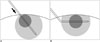
For index tumor located at superficial subcapsular region of the liver abutting the abdominal wall, the multi-tined expandable RF ablation electrode has the disadvantage that one or more of the tines may penetrate the liver capsule, resulting in injury of the abdominal wall during ablation. The cool-tip electrode has the disadvantage that the junction of insulated and exposed segment of the electrode cannot be detected by ultrasonography. Subsequently, the electrode may be positioned too deep within the liver parenchyma or may straddle the liver parenchyma and the abdominal wall. Moreover, even when the junction is placed exactly at the capsule of the liver, the ablation margin will be deviated toward the direction of electrode insertion, resulting in insufficient ablation margin in the proximal side.
The angled cool-tip electrode has the advantage that the electrode can be aligned in a symmetric configuration with respect to the index tumor and the liver capsule. In addition, the proximal end of the exposed segment of the electrode, which is the angle, can be detected by ultrasonography. As the angle is detectable, the index tumor can be centered within the exposed segment of the electrode. Subsequently, more favorable, symmetric ablation margin can be expected (Fig. 3). Minimal ablation margin of 5 mm is recommended in RF ablation of colorectal metastasis and 3 mm in hepatocellular carcinoma (222627). In using the angled cool-tip electrode for RF ablation of a superficial subcapsular tumor, a mean ablation margin of 7.07 ± 2.23 mm could be obtained without significant complication during mean follow up of 349.73 days.
Electrode tract seeding is another major delayed complication that can result after RF ablation of a subcapsular tumor as it results in distant metastasis, especially when performed as a bridge therapy for liver transplantation. The risk factors influencing electrode tract seeding include poorly differentiated tumor, high α-FP value in hepatocellular carcinoma, subcapsular location, tumor biopsy prior to RF ablation, numbers of sessions of treatment, numbers of electrode placement, tumor > 3 cm, direct subcapsular insertion of electrode, and intra-tumoral pressure increase (15161718). Decreasing the number of electrode insertion and repositioning, sufficient length of electrode tract, coagulation of electrode tract, and laparoscopic treatment of exophytic or subcapsular lesions are suggested to minimize the incidence of seeding (1117).
During RF ablation of a subcapsular tumor, it is difficult to acquire sufficient length of electrode tract during targeting and coagulation of the tract after the procedure. To avoid electrode tract seeding, direct penetration of the tumor without traversing non-tumorous liver parenchyma should be avoided (22), and laparoscopic RF ablation is recommended (1117). The alternative no-touch wedge ablation technique is reported for microwave ablation of subcapsular tumors in liver. The technique consists of placing the antenna in multiple sites in oblique directions tangential to the index tumor, without directly entering the tumor. The summated ablation zones are required to encompass the entire subcapsular tumor. But the technique has the disadvantage that some learning curve may be required as multiple ablation zones have to be designed to encompass the entire tumor. Occasionally, design of the ablation zones can be limited by intervening ribs or adjacent heat sensitive organs according to the tumor location. Requirement of multiple probe insertions with a mean of 5.6 insertions is another disadvantage of the technique (23). The technique of inducing artificial ascites or CT-guided targeting from distant access site to traverse sufficient length of liver parenchyma may also be used for targeting the superficial subcapsular tumor. But inducing artificial ascites requires additional time and procedure of catheter insertion, and may not be always successful, especially in patients who had undergone prior surgery. CT-guided targeting from distant access site has the disadvantage of radiation exposure and considerable amount of additional time may be required as the electrode has to pass through a lengthy tract, which may not be always easy.
The angled cool-tip electrode prevents direct penetration of the superficial subcapsular tumor, increases the length of electrode tract within non-tumorous liver parenchyma and ablates the electrode tract after ablation. Although the optimum length of electrode tract to minimize seeding is unknown, electrode tract seeding was not detected after RF ablation of subcapsular tumors using the angled cool-tip electrode during a mean follow-up of 349.73 days.
In preparing the angled cool-tip electrode, multiple small bends were made in as short length of the electrode as possible at the junction of insulated and exposed segment to avoid unnecessary exfoliation of the insulation. In addition, multiple small bends were added to angle the electrode rather than a single large bend to avoid fracture of the electrode while adjusting the tip of the electrode toward the epicenter of the tumor. In all our cases, no significant change in the angle of the electrode could be perceived after the procedure. Also, during test perfusion of the perfusate prior to the procedure, there was no leakage of the perfusate. Compared to using non-angled cool-tip electrode, there was no change in the total amount of perfusate used during the procedure and 1200 to 1500 mL of perfusate was used during 12 minute ablation.
We recommend bending the electrode contralateral to the attachment of tubes for perfusate, as in the transjugular intrahepatic portosystemic shunt needle (Fig. 1). Although an experienced interventional radiologist skilled in ultrasonography may perform the targeting without difficulty, bending the exposed electrode tip in a predictable direction would further ease the procedure. In addition, the weight of the tubes and the cable may rotate the electrode during ablation. In our experience, bending the electrode contralateral to the tubes could minimize the effect of weight of the tubes in most cases. Lastly, as the critical steps of the procedure are performed under ultrasonography guidance, high resolution ultrasonography equipment is recommended during targeting and monitoring.
The limitations in the study include its retrospective nature, small number of cases that were enrolled from a single institution, only small tumors included in the study, lack of histological confirmation of the tumor, and limited duration of clinical and imaging follow-up. Subsequently, the true recurrence rate and clinical significance of the technique are not yet known. Long-term follow-up in larger series of patients is required.
In conclusion, using an angled cool-tip electrode for RF ablation of superficial subcapsular tumor may be a feasible technique for adequate ablation margin and lowered incidence of complications. A larger prospective study is needed for a more definitive evaluation of the technique.
 XML Download