Abstract
Patients with Klippel-Feil syndrome (KFS) have an increased incidence of vascular anomalies as well as vertebral artery (VA) anomalies. In this article, we presented imaging findings of a 15-year-old female patient with KFS with a rare association of extraforaminal cranially ascending right VA that originated from the ipsilateral carotid bulb. Trifurcation of the carotid bulb with VA is a very unusual variation and to the best of our knowledge, right-sided one has not been reported in the literature.
Klippel-Feil syndrome (KFS) is a congenital spinal malformation associated with developmental anomalies of skeletal, neurological, cardiovascular and genitourinary system components. It was firstly described by Maurice Klippel and Andre Feil in 1912, typically with clinical triad including a short neck, low posterior hairline and limited range of motion. It is reportedly present in one of 42000 individuals and has female preponderance (57–70%) (12). Vascular anomalies such as anomalies of the aortic arch, carotid arteries, subclavian artery, vertebral artery (VA), and a persistent trigeminal artery have a increased incidence and are known in association with KFS (3).
Anomalous origin of VA is not a very common finding with incidence ranging from 3–8% (4). Multiple variations of the VA origin have been reported in the literature, including arising from the aortic arch, from the common, internal, or external carotid arteries and subclavian branches. However, anomalous origin of VA directly from carotid bulb is very rare.
In this article, we presented imaging findings of a 15-year-old female patient with KFS with an exceptional association of previously unreported extraforaminal cranially ascending right VA, which was originated from ipsilateral carotid bulb forming trifurcation appearance of right common carotid artery.
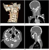
A 15-year-old female patient with known history of KFS including complaints of neck pain and occipital headache and dizziness that lasts for several months, was admitted to our hospital. Physical examination revealed reduced neck movement. Neurological examination and laboratory tests were normal. Radiographic studies depicted vertebral fusion in C2–4 level and right cervical tilt. No craniovertebral junction instability was detected in dynamic radiographic examination. Computed tomography (CT) revealed C1 right neural arch aplasia, hypoplasia of the right lateral mass of C2 and dysplasia of the right occipital condyle. In order to further clarify the presenting symptoms, craniocervical magnetic resonance imaging (MRI) were performed, showing absence of right VA at the foraminal level. Spinal cord compression was absent. Due to the MRI findings, cervical CT angiography examination was done and revealed absence of right VA in transverse foramina. Additionally to external and internal carotid arteries, the right VA was also originated from the carotid bulb (trifurcation). The anomalous VA, without entering the transverse foramina, ascended cranially in the prevertebral space and entered the cranium through the foramen magnum anteriorly (Fig. 1A-D). Except for C7 neural foramen (5 mm), neural foramina of C6 and above were hypoplasic (2–3 mm). Although the leftside neural foramina were in normal calibration, left VA was hypoplastic.
Anomalies concomitant to KFS include spinal abnormalities (hemivertebrae, spina bifida, cervical diastematomyelia, anterior vertebral body cleft, and butterfly vertebrae) (1), skeletal system abnormalities (scoliosis and/or kyphosis [60%], Sprengel's deformity [30%], and torticollis), urinary system abnormalities (35%), loss of hearing (30%), facial asymmetry and flattening of the neck (20%), synkinesis or mirror movements (20%) and congenital heart disease (4% to 14%) (3). Patients with KFS show increased incidence of vascular anomalies. Understanding this concomitancy requires knowledge of embryology. During the fifth week of gestation, a transverse fissure divides the cranial and caudal halves of these sclerotomes. Failure in segmentation of the intervertebral disks may result in KFS. Vascular system development parallels the development of the spinal cord (1). At the 7-mm stage of embryonic development, seven cervical intersegmental arteries (CIAs) appear. At the 14- to 17-mm stage, the horizontal parts of the first six CIAs disappear on involution and the remaining 7th CIA becomes part of the subclavian artery. The VA typically originates from the distal end of the 7th dorsal intersegmental artery bilaterally and with the persistance of longitudinal anastomosis. Failure of involution (or a persistance of CIA) in one of the first six CIAs, depending on the CIA level, causes the developement of anomalous origin of VA (Fig. 2) (5).
Vasović et al. (6) described that primitive carotid-vertebrobasilar anastomoses via the primitive proatlantal intersegmental arteries (PPPA) provide the proximal supply to the longitudinal neural arteries (6). PPPA is divided into two types. Type 1 PPPA originates from the internal carotid artery or the external carotid artery (ECA) at the proximal portion, and ascends posterolaterally. It runs without passing through the transverse foramen of the cervical vertebrae, and joins the intracranial VA after running through the foramen magnum (as in our case). Type 2 PPPA arising from the ECA ascends more laterally and runs through the transverse foramen of C1 vertebra and then joins the extracranial VA below the C1 level (7). In our case, probable persistence of right PPPA remaining as right VA with obliteration of proximal longitudinal anastomoses constitues the possible embryological development of the anomalous origin of right VA from right carotid bulb.
Anomalous origin of left VA is more frequently involved than the right. Origin of left VA directly from aortic arch (about 7% of cases), represents the commonest variation in the origins of VAs and second commonest aortic arch anomaly (8). However, anomalous origin of VA directly from carotid arteries is very rare. VA originating from common carotid artery (5) and from ECA (9) have been reported in the literature. Trifurcation of common carotid artery is well known and trifurcation with occipital artery and facial artery have been reported (10). However, carotid trifurcation with VA is an unusual condition and the left-sided variety was firstly reported by Patil et al. (4). They reported imaging findings of acute infarct in ipsilateral thalamus of a male patient in whom trifurcation of left common carotid artery was depicted. Regarding embryological development, right-sided carotid trifurcation with VA is likely as relevant as previously reported left-sided cases and, to the best of our knowledge, has not been reported in the literature.
Normally, both VAs arise from subclavian artery, except for C7 transverse foramen, they pass through C6 to C1 transverse foramina and enter foramen magnum with its intracranial course. Importantly, in our case, abnormal course of right VA occured after originating from carotid bulb. Patil et al. (4) reported a case of anomalous left VA that originated from carotid bulb as trifurcation, and subsequently entered left transverse foramen of C1. Above C1 level and also intracranial course of VA was noted as normal in their case. However in our case, the right VA originated from the right carotid bulb, without passing through any transverse foramen, ascending at the anterior aspect of the cervical vertebrae, eventually entering the foramen magnum anteriorly just close to the clivus.
In cases with anomalous arterial anatomy, alterations in hemodynamics may occur and may lead to cerebrovascular accidents or predispose to aneurysms due to turbulence. Also, the carotid bifurcation is the most common site of atheromatous plaques. In cases of carotid trifurcation with VA, occlusion of vertebral/basilar artery may occur by the embolic sources from carotid bulb and may result in ischemic symptoms in posterior cerebral territories (6). Additionally, regarding anatomic location of anomalous VA, pathologies in cervical and craniocervical junction may cause similar vascular problems. Also, the concomitancy of cervical congenital anomalies, such as KFS, and vascular anomalies comprise high risk for vascular injury during surgery. Preoperative imaging and further clarifying of vascular structures is helpful to reduces the risk of surgical vascular complications.
Figures and Tables
 | Fig. 13D CT demonstration of anomalous right VA.3D volume rendered CT (A), coronal (B), axial (C), and sagittal (D) maximum intensity projection CT images show right vertebral artery originating from carotid bulb. Internal carotid artery (white arrow in A and B), external carotid artery (black arrows in A), vertebral artery (arrowheads in A and B). Images show course of right vertebral artery (arrows in C and D) in prevertebral location, its ascending in foramen magnum anteriorly without entering any transverse foramen. Right and left internal carotid arteries (arrowheads in C). VA = vertebral artery, 3D = three-dimensional
|
 | Fig. 2Schematic illustration aortic arch system.
A. Normal embryologic development. Horizontal parts of first six CIAs disappear on involution, bilateral regression of DC separates common carotid arteries from subclavian artery on right side and from aortic arch on left side. Aortic arches (I–VI) are illustrated. PPPA provide proximal supply to longitudinal neural arteries. B. Normal anatomy in adults: longitudinal connections linking intersegmental arteries that further form VA bilaterally from ipsilateral subclavian arteries, which are derived from seventh intersegmental arteries. AoArch = aortic arch, BA = basilar artery, CCA = common carotid artery, CIAs = cervical intersegmental arteries, DC = ductus caroticus, ECA = external carotid artery, ICA = internal carotid artery, LScA = left subclavian artery, PPPA = primitive proatlantal intersegmental arteries, RScA = right subclavian artery, VA = vertebral artery
|
References
1. Ulmer JL, Elster AD, Ginsberg LE, Williams DW 3rd. Klippel-Feil syndrome: CT and MR of acquired and congenital abnormalities of cervical spine and cord. J Comput Assist Tomogr. 1993; 17:215–224.
2. Guille JT, Sherk HH. Congenital osseous anomalies of the upper and lower cervical spine in children. J Bone Joint Surg Am. 2002; 84-A:277–288.
3. Sudhakar AS, Nguyen VT, Chang JB. Klippel-Feil syndrome and supra-aortic arch anomaly: a case report. Int J Angiol. 2008; 17:109–111.
4. Patil PV, Patil AM, Apte AV, Attarde VY. Anomalous origin of left vertebral artery from carotid bulb seen as "trifurcation" of left common carotid artery with acute infarct in ipsilateral thalamus: a case report. J Neuroimaging. 2015; 25:662–664.
5. Chen CJ, Wang LJ, Wong YC. Abnormal origin of the vertebral artery from the common carotid artery. AJNR Am J Neuroradiol. 1998; 19:1414–1416.
6. Vasović L, Mojsilović M, Andelković Z, Jovanović I, Arsić S, Vlajković S, et al. Proatlantal intersegmental artery: a review of normal and pathological features. Childs Nerv Syst. 2009; 25:411–421.
7. Woodcock RJ, Cloft HJ, Dion JE. Bilateral type 1 proatlantal arteries with absence of vertebral arteries. AJNR Am J Neuroradiol. 2001; 22:418–420.
8. Welch CS, Stark P. The five-vessel arch: independent origin of both vertebral arteries from the aortic arch. J Comput Assist Tomogr. 2012; 36:275–276.
9. Nasir S, Hussain M, Khan SA, Mansoor MA, Sharif S. Anomalous origin of right vertebral artery from right external carotid artery. J Coll Physicians Surg Pak. 2010; 20:208–210.
10. Altinbas NK, Unal S, Peker A, Uzun C, Akkaya Z, Peker E. Trifurcation of the left common carotid artery. Pol J Radiol. 2015; 80:376–378.


 PDF
PDF ePub
ePub Citation
 Print
Print


 XML Download