

 PDF
PDF ePub
ePub Citation
 Print
Print


INTRODUCTION
Epidural steroid injections (ESIs) are frequently employed for the conservative management of low back pain caused by spinal stenosis or herniated intervertebral discs. Without fluoroscopy, inappropriate needle placement has been reported in as many as 30% of ESIs, even with experienced practitioners (12). Fluoroscopy ensures the accuracy of therapeutic injections, with a sufficient percentage of ESIs reaching the target site, and thus, could affect treatment outcomes (3456). This technique may reduce the complications associated with ESIs, such as intravascular injections, dural punctures, paraplegia secondary to spinal cord infarctions, cerebellar infarctions, and even death (678910).
Fluoroscopy has been used in at least 50% of interventions in the area of pain management, including ESIs, and its use has grown considerably (1112). The increase in fluoroscopy-guided ESIs has led to a growing concern of radiation exposure and radiation dose. In order to mitigate potential radiation hazards, multiple techniques have been introduced, including protection, decreasing the duration of the intervention, very low frame rate-pulsed fluoroscopy, removal of the grid, last image hold and electronic collimation, and intermittent fluoroscopy (13141516).
For ESIs in the lumbar spine, continuous fluoroscopic monitoring (CM) is employed during needle advance to ensure correct needle positioning. In our opinion, CM during needle advance has several advantages over intermittent fluoroscopic monitoring (IM). Firstly, CM is thought to be safer for patients during needle advance because the needle can be seen directly. Secondly, it can reduce intervention time, due to lower likely hood of need for needle repositioning. Thirdly, it is useful for trainees who are learning the procedure and have less experience and skill with ESI techniques than experienced practitioners. The main disadvantage of CM is that it involves a higher risk of radiation exposure. To our knowledge, there have been no reports on the use of CM during lumbar ESIs in terms of radiation exposure and intervention time, although there have been several reports on radiation exposure during spine injection with IM.
We hypothesized that although CM during needle advance in ESIs leads to higher radiation exposure, this exposure will be within tolerable limits, because no increase in intervention time will be required, even for trainees. The purpose of this prospective study was to evaluate radiation exposure (dose area product [DAP] and fluoroscopy time) and intervention time during lumbar ESIs under CM by analyzing the differences among the various practitioners (two experienced radiologists and a trainee) and among different ESI methods. In addition, we aimed to compare radiation exposure and intervention time between CM and IM in procedures undertaken by one senior faculty member.
MATERIALS AND METHODS
Patients
Patients who underwent lumbar ESIs for radiculopathic low back pain in a tertiary university teaching hospital from July 2011 to October 2011 were consecutively recruited. All patients provided written informed consent to the study. This study was reviewed and approved by our Institutional Review Board for human investigation.
Treatment
Lumbar ESIs were performed under fluoroscopic guidance by three physicians (a senior faculty member who had conducted > 10000 ESI procedures over 10 years, a junior faculty member who had performed > 5000 ESI procedures over 5 years, and a trainee in the process of fellowship) employing different methods (caudal, interlaminar, and transforaminal ESIs). The level of injection varied according to the patients' symptoms and the location of the nerve root compression.
Only the senior faculty member used both fluoroscopic monitoring methods: CM was performed before September 1, 2011, and IM was carried out thereafter. The IM method involves short-duration radiation exposure, which is sufficient to view the position of the needle; whereas in the CM method, radiation exposure occurs for the entire duration of the intervention. The other two physicians (the junior faculty member and the trainee) used CM in all patients.
All injections were performed with patients in the prone position using one of two angiography units randomly: the two uniplanar digital subtraction angiography units (Integris Allura Xper FD 20; Philips Healthcare, Best, the Netherlands) or the biplanar digital subtraction angiography unit (Intergris Xper FD 20; Philips Healthcare, Best, the Netherlands). During caudal ESI, a 22-gauge spinal needle was inserted through the sacral hiatus and advanced into the sacral canal to the S3 level. For transforaminal ESI, a 22-gauge spinal needle was advanced into the so-called "safe triangle," which is defined superiorly by the pedicle, laterally by the lateral border of the vertebral body, and medially by the outer margin of the spinal nerve (17). Subsequently, contrast media (Omnipaque 300 [iohexol, 300 mg iodine per mL]; Amersham Health, Princeton, NJ, USA) was injected to confirm the needle's placement in the epidural space. For interlaminar ESI, a 22- or 25-gauge spinal needle was inserted into the posterior epidural space until the contrast media smoothly spread into the lumbar epidural space (17). Additional details of each method are provided in our previous report (18). Fluoroscopic images were obtained during these procedures, in order to check the accuracy of the needle position. Subsequently, a mixture of triamcinolone acetonide (Triam 40 mg/1 mL; Shin Poong Pharmaceutical, Seoul, Korea) was injected into the epidural space, followed by injection of the anesthetic mixture, including bupivacaine hydrochloride (0.5 mL/0.5%, Marcaine Spinal 0.5% Heavy; AstraZeneca, Westborough, MA, USA). All physicians used standard radiation protection in accordance with the as low as reasonably achievable (ALARA) principle recommended by the International Commission on Radiological Protection (19).
Assessments
Fluoroscopy time during intervention and DAP as a radiation dose to which patients were exposed were automatically measured and recorded by the uniplanar or biplanar digital subtraction angiography units. In addition, intervention time was documented manually from the beginning to the end of each intervention by one of two researchers. The beginning and end points were defined as when the needle penetrated the skin and when it was removed from the skin, respectively.
Statistical Analysis
With CM, a three (practitioner: a senior faculty member, a junior faculty member, and a trainee) by three (ESI methods: caudal, interlaminar, and transforaminal ESI) ANOVA was conducted on DAP, fluoroscopy time, and intervention time to determine the single effects of the variables and their interaction. A two-way ANOVA with interactions was used to determine the effect of the monitoring method (continuous vs. intermittent) and different methods by a senior faculty member. For all statistical tests, a p value < 0.05 was considered significant. All statistical analyses were performed using SPSS 17.0 (PASW, version 17.0; SPSS Inc., Chicago, IL, USA).
RESULTS
Participants
A total of 804 patients (520 females and 304 males) with a mean age of 64.1 ± 13.7 years (18–89 years) were enrolled. Thirty patients were excluded because of errors in recording the radiation dose and intervention time and 14 patients whose practitioners were promoted from trainee to faculty member during interventions were also excluded. One patient whose intervention was converted from a caudal to an interlaminar ESI following the unexpected finding of a Tarlov perineural cyst was excluded. No adverse events or complications occurred during the interventions.
As a result, 759 patients (472 females and 287 males, mean age: 64.0 ± 13.7 years) who underwent 922 interventions (double and triple procedures: 119 and 22 patients, respectively) during the study period were investigated. A total of 824 and 98 interventions were performed with the uniplanar and biplanar digital subtraction angiography units, respectively. The employed units were similar between practitioners (χ2 = 1.99, p = 0.369) and different ESI methods (χ2 = 3.02, p = 0.221) with continuous monitoring, and between monitoring methods (χ2 = 0.03, p = 0.862).
Subjects were divided into 3 groups based on the physician performing the interventions (a senior faculty member, a junior faculty member, and a trainee), and the senior faculty member group was further divided into two groups according to the monitoring method. Demographic data relating to age, gender, number of patients, and number of interventions according to the physician were shown in the Table 1.
Radiation Exposure and Intervention Time According to Practitioners and Interventions during Continuous Monitoring
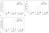
Figure 1 illustrated the mean DAP, fluoroscopy time, and intervention time according to ESI methods for each practitioner with CM. Simple main effects analysis showed that DAP differed significantly according to practitioner (F [2, 724] = 26.180, p < 0.001) and ESI methods (F [2, 724] = 52.702, p < 0.001), but that there was a significant interaction between the effects of the practitioner and the ESI methods on DAP (F [4, 724] = 8.791, p < 0.001). Post hoc Tukey's tests showed that DAP was lower in more experienced practitioners (the senior and junior faculty members) than in the less experienced trainee (p < 0.001), although it was similar between the faculty members (p = 0.944). In addition, post hoc comparisons showed that DAP was lower in transforaminal ESIs, as compared to interlaminar and caudal ESIs, and lower in interlaminar ESIs, as compared to caudal ESIs (p < 0.001).
There was a significant main effect according to practitioner (F [2, 724] = 31.792, p < 0.001) and ESI methods (F [2, 724] = 12.523, p < 0.001) on fluoroscopy time without any correlation between practitioner and intervention (F [4, 724] = 0.588, p = 0.672). Post hoc Tukey's tests showed that fluoroscopy time was shorter in the faculty members than in the trainee (p < 0.001) and was similar between the faculty members (p = 0.586). Post hoc Tukey's tests showed that fluoroscopy time was longer in interlaminar ESIs, as compared to caudal and transforaminal ESIs (p < 0.001). However, fluoroscopy time was similar between caudal and transforaminal ESIs (p = 0.842).
Significant main effects of the practitioner (F [2, 724] = 12.738, p < 0.001) and ESI method (F [2, 724] = 19.525, p < 0.001) on intervention time were indicated without correlation between the practitioners and ESI methods (F [4, 724] = 0.339, p = 0.852). A post hoc Tukey's test showed that intervention time was shorter in the faculty members than in the trainee (p < 0.001) and similar between faculty members (p = 0.144). Additionally, post hoc Tukey's tests showed that intervention time was longer in interlaminar ESIs, as compared to caudal (p = 0.001) and transforaminal ESIs (p < 0.001), and longer in caudal ESIs, as compared to transforaminal ESIs (p = 0.005).
Radiation Exposure and Intervention Time According to Monitoring Methods and Interventions in a Senior Faculty Member
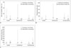
Figure 2 depicted the mean DAP, fluoroscopy time, and intervention time according to ESI methods with two different monitoring methods (continuous and intermittent) in a senior faculty member. Simple main effects analysis showed that DAP with IM was significantly lower than that with CM (F [2, 328] = 99.549, p < 0.001); and that DAP was different according to ESI methods (F [2, 328] = 20.172, p < 0.001). However, there was no difference between DAP of transforaminal ESI with CM and that of caudal or interlaminar ESI {interaction between the monitoring methods and ESI methods on DAP (F [2, 328] = 10.477, p < 0.001)}. DAPs for caudal, interlaminar, and transforaminal ESIs were 1.19 ± 1.40, 1.14 ± 0.75, and 0.73 ± 0.83 Gy·cm2 with IM, in contrast to 5.26 ± 3.76, 3.90 ± 5.03, and 2.19 ± 1.67 Gy·cm2 with CM, respectively. There was a significant main effect of the monitoring method (F [2, 328] = 68.261, p < 0.001) and ESI method (F [2, 328] = 5.648, p = 0.004) on fluoroscopy time, without any interaction between monitoring method and intervention (F [2, 328] = 1.771, p = 0.172).
A significant main effect of the intervention on intervention time (F [2, 328] = 10.266, p < 0.001) was shown, but there were no differences on intervention time between monitoring methods (F [2, 328] = 0.432, p = 0.511), without interaction between the monitoring method and ESI method (F [2, 328] = 1.095, p = 0.336).
DISCUSSION
Intermittent monitoring involved substantially lower DAP and less fluoroscopy time, without a difference in the intervention time, as compared to CM in a senior faculty member. IM is reportedly effective in reducing radiation exposure (1315). However, no study has actually demonstrated that IM results in a lower radiation dose, as compared to CM. Furthermore, few studies have investigated the degree to which radiation can be reduced by using IM in comparison to CM.
Our results showed that DAPs for caudal, interlaminar, and transforaminal ESIs were 4.4, 3.5, and 3.1 times higher with CM than with IM, respectively in a senior faculty member. After the International Commission on Radiological Protection first provided reference-level values in 1991 (20), the diagnostic reference levels have been used as a norm in order to avoid unnecessary radiation risks. Additionally, in fluoroscopy-guided interventions, the reference levels could be applied to protect the patients or occupationally exposed staff from radiation. A report on 26 radiation-induced skin injuries due to fluoroscopy-guided interventions highlights the need to consider the radiation dose level when using fluoroscopic guidance (21). However, establishing the reference levels in the area of fluoroscopic intervention is difficult because of the variability of the duration and complexity of each intervention. Therefore, radiation exposure should be minimized, in keeping with the ALARA philosophy in pain intervention (22). Thus, our results suggested that IM is preferable to CM in terms of radiation exposure dose in an experienced practitioner. Additionally, our study showed that CM resulted in 3.0, 3.1, and 2.7 times more fluoroscopy time than IM in caudal, interlaminar, and transforaminal ESIs, respectively. However, there was no significant difference in intervention time between CM and IM. In summary, IM seems preferable to CM in terms of radiation safety without a delay in intervention time.
However, adverse events occur more frequently with IM-guided pain intervention. A variety of complications secondary to interventional pain management, ranging from minor to severe complications, such as paraplegia, epidural and subdural hematomas, and even death, have been documented (8232425). These adverse events are thought to arise from incorrect needle positioning, and several methods have been employed to avoid them (26). CM has been recommended as one such method; however, it is not as commonly used as IM, despite the benefit of avoiding intravascular injection. This recommendation is especially important for less experienced practitioners, who lack a thorough understanding of the procedure, relevant anatomy, and fluoroscopic imaging. Under these circumstances, CM is favored, because decreased procedure-relevant risk might be greater than the multiplied risk from an additive radiation dose exposure. Thus, only the most experienced senior faculty member utilized IM in our study. However, how much experience is required to engage in IM effectively without raising concerns regarding intervention-related complications remains unclear and requires further research.
Because a comprehensive assessment of both deterministic effects and stochastic aspects is critical to evaluating the potential risk of radiation, both the entrance surface dose and effective dose of radiation to the patient should be obtained (1520). In terms of deterministic effects, skin injuries have been a major concern in fluoroscopy-guided intervention (2327). However, the direct measurement of radiation dose to the skin and of the effective dose is not straightforward in clinical practice; thus, they may be estimated using conversion factors from DAP instead of direct measurement. In the absence of specific conversion factors for interventional pain management, we adopted a published conversion factor from cardiac intervention and used the highest value in order to verify the radiation effect, 9.7 mGy/Gy·cm2 for the skin dose and 0.27 mSv/Gy·cm2 for the effective dose (2728). Applying the same conversion factor to the highest DAPs from the present study, i.e., the caudal ESIs performed by the trainee, a 546.34 mGy of radiation dose and 15.21 mSv of effective dose were calculated. The suggested threshold for radiation-induced skin injury is 2 Gy and the annual exposure allowance limit is 50 mSv (1929). Therefore, lumbar ESIs under CM were within tolerable limits in terms of potential risk of radiation to the patients, even when performed by the trainee. However, radiation hazard to the radiologists who performed the interventions may be a serious concern, even though protective garments such as lead aprons, lead glasses, and radiation attenuation gloves are used. A recent study reported radiation-induced hand necrosis in a physician who had performed fluoroscopy-guided spine injections for 17 years (30). Therefore, medical staff, including radiologists, should be aware of the hazards of radiation and make efforts to minimize radiation exposure.
Fluoroscopy time was utilized to compare our results to those of previous studies, because of a lack of relevant studies using DAP as a radiation dose in pain intervention. The exposure time in our study was longer than that reported by Manchikanti et al. (1331) but similar to that of Botwin et al. (3233). This disparity is most likely due to the different measurement methods; thus, a direct comparison was not possible. However, a similar discrepancy between different ESI methods, which commonly occurred in previous studies, indicates that radiation dose exposure depends on the ESI methods. In addition, the physician's level of experience was found to be one of the factors affecting radiation exposure, consistent with the results of a previous study (13).
Our study has several limitations. IM was used by the senior faculty member alone; thus, comparisons between IM and CM were not generalized to the other physicians. However, we hypothesized that the risk of complications secondary to incorrect needle positioning should not be ignored in inexperienced practitioners in order to protect the safety of subjects. In addition, this study was not randomized; the participants were consecutively recruited and allocated. Because of the lack of previous direct comparisons between different practitioners and monitoring methods, we were unable to demonstrate the safety or efficacy of this study. The results of the present study could form the basis for further advanced studies with randomization.
In conclusion, radiation dose exposure during lumbar ESIs under CM was different according to the practitioners and methods and within the established safety limits. For a senior faculty member, IM resulted in substantially less radiation exposure, as compared to CM during lumbar ESIs. Considering the uncertain cumulative, long-term effects of radiation, for experienced practitioners, IM might be more desirable in order to keep radiation dose exposure within the ALARA principle. However, in view of the complications during IM-guided pain intervention other than radiation exposure, CM should be considered for inexperienced trainees.

 XML Download