

 PDF
PDF ePub
ePub Citation
 Print
Print


INTRODUCTION
Pulmonary developmental anomalies are considered to span a continuum, ranging from an abnormal lung containing normal vessels to a normal lung containing abnormal vessels (1). Although many cases of pulmonary developmental anomalies have the classic features of a single anomaly, other cases have features of two or more anomalies. We encountered a unique case of a patient with segment of a hyperlucent lung with no normal bronchial connection and an aberrant pulmonary arterial supply, which does not match any other previously reported category. This case report was approved by our Institutional Review Board and the requirement for the patient's informed consent was waived.
CASE REPORT
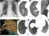
A 64-year-old man who was experiencing mild chest discomfort visited our hospital. His physical examination was unremarkable and there was no significant medical history. He underwent routine chest radiography (Fig. 1A) that showed an area of hyperlucency at the right lower lung zone, but no air-containing cysts or any other abnormal opacity.
The multidetector computed tomography scan was performed on a 64-slice CT system (Discovery CT 750HD; GE Healthcare, Milwaukee, WI, USA). Precontrast and postcontrast CT scans were performed with iodinated non-ionic contrast, 90 cc/3 cc/s, injected using a pressure injector. Then, a series of inspiratory and expiratory high-resolution CT (HRCT) scans were obtained at 0.6-mm collimation and 1-cm intervals. CT showed an area of increased lucency at the right lower medial lung, and a tortuous, ectopic bronchus with bronchiectasis was identified within the hyperlucent area (Fig. 1B); furthermore, communication between the ectopic bronchus and the right bronchial tree was absent, and the right medial basal segmental bronchus was obliterated (Fig. 1C). The ectopic bronchus communicated with no other structures such as the esophagus or mediastiunum, but showed communication with a peripheral bronchiolectasia that ran upward and had a bubbly appearance (Fig. 1B). The mediastinal setting in the CT scan revealed an anomalous pulmonary artery arising from the posterior inferior aspect of the right main pulmonary artery and supplying the affected region. A three-dimensional volume-rendering image was reconstructed to properly demonstrate the abnormal pulmonary artery (Fig. 1D). The segmental vein drained into the right pulmonary vein.
Expiratory HRCT scans were obtained in an attempt to show air trapping within the abnormally lucent lung. These images showed numerous, moderately thin-walled, air-containing cysts, 0.5 to 1.0 cm in diameter that were mainly located in the peripheral zone of the hyperlucent segment (Fig. 1E). Several areas within the lucent segment that contained small cysts showed an increase in attenuation density during exhalation, but this increase had a delayed rate and a lesser degree, than the density increase seen in the normal contralateral lung (Fig. 1E). The patient's symptoms improved and he refused to undergo further evaluation.
DISCUSSION
The two major components of this case were 1) hyperlucent aeration of a sequestered segment with an independent ectopic bronchus that had no connection with the other bronchus or organs such as the esophagus and 2) an aberrant pulmonary artery from the right pulmonary trunk supplying the involved lung segment. These findings indicated a continuum of maldevelopment involving the pulmonary parenchyma or the pulmonary vessels, or a combination of both; this spectrum has been called the "sequestration spectrum" (1). Thus, this case represents a spectrum of congenital bronchopulmonary malformations that has not been reported previously.
In the present case, thin section CT revealed proximal obliteration of the medial basal segment of the right lung, which is a feature of bronchial atresia. However, there was no evidence of mucocele or bronchocele at the affected segment, as is frequently seen in bronchial atresia; thus, this case was distinguished from bronchial atresia. A tortuous, bronchiectatic ectopic segmental airway was identified within the affected segment. Similar findings were reported in a pulmonary sequestration report (2).
On lung window images, CT clearly showed that the medial basal segment of lung lacked normal communication with the tracheobronchial tree or any adjacent organ or mediastinum. However, the manifested segment appeared as an area of hyperlucency that remained at the end of forced expiration, confirming air trapping. The mechanisms by which the segment was aerated are unclear. Stern et al. (3) suggested that aerated pulmonary sequestration is caused by collateral pathways between the medial basal segment and the right lung through pores of Kohn that are probably located at the level of the lung parenchyma, as might also be applicable to the current case. In a review on the CT appearances of cases with aerated bronchopulmonary sequestration (4), the presence of lucentor low-attenuation areas of "emphysema" is emphasized. The emphysema in patients with sequestration is suggested to result from collateral ventilation and air trapping (5). In our case, the emphysematous small cystic feature within the involved segment is clearly seen on HRCT, which can be a potential collateral aeration channel. The location of emphysema or aircysts, which were found mainly at the peripheral zone and adjacent to the normal lung, also supports this idea. Furthermore, the ectopic bronchus is possibly aerated by a small airway connected to peripheral bronchiolectasia, as in our case. On CT, the ectopic bronchus, ran upward and tapered into bronchiolectasia with an unusual bubbly appearance. This unexpected finding could be a clue to collateral aeration channel in addition to the peripheral collateral ventilation through pores of Kohn.
This patient also showed an aberrant pulmonary artery from the right pulmonary trunk supplying the involved lung segment. This is a discrepant feature, since most vascular anomalies in the "sequestration spectrum" are supplied by aberrant systemic arteries. Moreover, in reviewing 524 lungs or lobes of resection performed on 426 patients, Cory and Valentine (6) described no variation of the pulmonary artery in the primary lung hilum. However, our case showed early branching of a segmental pulmonary artery before the right main pulmonary trunk entered the lung hilum. A similar vascular abnormality has not been reported previously, to the best of our knowledge. If overlooked, anatomical variations of the lung vessels may be important obstacles during lung surgery. This case suggested pulmonary artery abnormality at the hilar level. Moreover, in early fetal development, the airways appear to act as a template for pulmonary artery development and central pulmonary artery sprouting or angiogenesis for up to approximately seven generations (counting the artery to each lung as the first generation) (78). The findings in our case were suggestive of a unique combination of congenital airway and vascular anomalies, rather than an acquired lesion.
In summary, we presented a case of combined anomalies of a hyperlucent lung segment supplied by ectopic bronchus and an aberrant pulmonary artery. Our patient is currently receiving regular expectant follow-up, with particular attention to infection or impaired pulmonary function.
 XML Download