

 PDF
PDF ePub
ePub Citation
 Print
Print


INTRODUCTION
Uterine intravenous leiomyomatosis (IVL), an unusual growth pattern of uterine leiomyoma, is a rare neoplasm characterized by intravascular proliferation of a histologically benign smooth muscle cell tumor. IVL could act in a somewhat malignant fashion, extending along the pelvic veins, adrenal or renal veins to the inferior vena cava (IVC), occasionally reaching as far as the right cardiac chambers and the main pulmonary artery, or producing benign metastases, usually to the lungs or lymph nodes (12).
Herein, we reported an interesting case of a patient suffering from endovascular leiomyomatosis of the uterus with pulmonary metastases and extension to the right cardiac chambers. To our knowledge, this was the first report of fluorodeoxyglucose (FDG) positron emission tomography/computed tomography (PET/CT) imaging findings in an IVL patient with involvement of lungs as well as cardiac chambers.
The report was approved by the Institutional Review Board of Gansu Provincial Hospital.
CASE REPORT
A 48-year-old previously healthy woman with a 50-day history of irregular vaginal bleeding and intermittent abdominal pain was transferred to our hospital for further evaluation. The patient had no previous surgery or trauma history. Physical examination on admission showed no abnormal finding. Laboratory studies revealed a decreased hemoglobin count (96 g/L; normal range 110–150 g/L). The levels of serum tumor markers, such as alpha-fetoprotein, carcinoembryonic antigen, squamous cell carcinoma antigen, CA19-9, CA125, and CA72-4 were normal. A pelvic ultrasound from another institution showed a giant solid mass in the pelvic cavity, and the possibility of gynecologic malignant tumor was raised.
18Fluorodeoxyglucose positron emission tomography-computed tomography (Discovery STE 16, General Electric Healthcare, Fairfield, CT, USA) scan was performed to further assess the nature of the pelvic abnormality and detect other abnormal lesions. The patient had a blood glucose level of 5.1 mmol/L on fasting for 6 hours prior to the study. CT and PET images were consecutively acquired from the top level of the skull to the middle thighs 1 hour after the injection of 259 MBq (7 mCi) of FDG. The acquired data were reconstructed using an iterative reconstruction and CT-based attenuation correction.
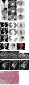
Except for the obvious physiological FDG uptake of colon and endometrium (Fig. 1A, arrows), the maximum intensity projection, axial CT and PET images shown in Figure 1A demonstrated low accumulation of FDG in the lobulated, aggressive occupying lesion in the abdominal and pelvic cavity, which was closely adjacent to the uterus and extended upward to the inferior of kidneys with a CT number of 20–50 Hounsfield unit and a size of 19.8 × 23.0 × 7.5 cm, approximately. The maximum standardized uptake value (SUVmax) was 1.6. In addition, multiple nodules in bilateral lungs without hypermetabolic foci were found (Fig. 1B, arrows); and the SUVmax and the size ranged from 0.5 to 1.0 and 0.6 × 0.8 cm to 1.7 × 2.1 cm, respectively. Furthermore, a hypodense lesion with mild FDG uptake was observed in the right ventricle (Fig. 1C, arrows), with the SUVmax of 2.1. Based on the FDG PET/CT findings, the presumptive diagnosis was IVL with intracardiac extension and pulmonary benign metastases.
Abdominal MRI combined with cardiac MRI was performed 1 day later. Abdominal enhanced MRI displayed intravascular filling defects in both enlarged ovarian veins (Fig. 1D, arrows), also in the IVC extending into the right atrium (Fig. 1E, arrow). MR white-blood sequence (Fig. 1F-H) showed three oval masses in the right cardiac ventricle (arrows); of which, a smaller mass (1.8 × 1.1 cm in diameter) moved back and forth through the tricuspid orifice (Fig. 1F, arrow), and the largest mass (2.6 × 2.8 × 3.8 cm in diameter) extended into the right ventricular outflow tract (Fig. 1H, arrow).
The patient then received a CT guided biopsy of the abdominal and pulmonary masses. The histopathological appearance of both lesions (Fig. 1I) showed interlaced bundles of spindle cells with homogeneous size, oval nuclei, eosinophilic cytoplasm, rare mitotic figures, and decorated by several thick-walled small blood vessels. These features were consistent with a leiomyoma, which further verified the diagnosis of IVL.
DISCUSSION
Intravenous leiomyomatosis, a condition that only affects women, is histologically confirmed benign smooth muscle tumor within vascular spaces from intrauterine venules to the systemic veins including iliac vein, IVC, even extending into the right heart chamber (12). Most IVL patients have a history of previous hysterectomy, or symptoms due to uterine fibroids (34). A review of the cases reported in the literature demonstrated that 64% of the women had undergone a previous hysterectomy ranging from 6 months to 20 years prior to presentation with the intravenous portion of the tumor (3). However, our patient had no history of surgery, but instead, a 50-day history of gynaecological symptom.
Intravenous leiomyomatosis is a very rare condition, thus it is usually diagnosed after an operation for myoma uteri. Its extrauterine involvement occurs in approximately 30% of cases and intracardiac extension accounts for about 10% (4). According to the reported literature (56), IVL with cardiac extension is relatively frequent, and IVL with pulmonary metastases is less common. Only one surgical case reported a combination of IVL with intracardiac extension and pulmonary benign metastases (7).
Besides surgery, imaging modalities including CT, MRI, or ultrasound could display different lesion profiles. However, due to the limitations of regional and anatomical imaging, it is relatively difficult to acquire systemic recognition and assessment of the nature of lesion. FDG-PET is based on the differential uptake of FDG by metabolizing cells and is transported into cells on the basis of their rate of glycolysis. PET scans can visualize active neoplastic lesions as areas of focal hypermetabolism (8). Therefore, FDG-PET is a sensitive modality for the diagnosis and staging of various malignancies. However, the characteristic of IVL on FDG PET/CT are unclear.
A few case reports have indicated that FDG PET/CT findings of benign metastasizing leiomyoma of the lungs include faint or non-avid metabolic activity of the nodules (910). Due to its similar histology with benign leiomyomata, IVL lesion also has a mild or absent FDG uptake on PET/CT images, as observed in our case. FDG PET/CT fusion imagery is a powerful tool for detecting systemic lesions. In our case, at the time of assessing the nature of pelvic primary tumor, FDG PET/CT could survey the entire body glucose metabolism and detect benign secondary lesions, including the right ventricle mass, and the metastasizing lung nodules.
According to previous reports, IVL with cardiac involvement is usually depicted on echocardiogram, contrast-enhanced CT or MRI and rarely detected with FDG PET/CT (12345). In the present case, FDG PET/CT showed only one of three masses in the right cardiac chambers explored by the cardiac MRI, while the intravenous lesions were invisible. MRI has an excellent soft-tissue resolution, whereas PET/CT has poor efficiency in the isodensity lesion with mild FDG distribution, as in the intravascular lesions presented in this case. Furthermore, the lesion size is also an important factor in the diagnostic imaging, therefore the largest mass in the cardiac cavity was revealed on FDG PET/CT. Consequently, whole body FDG PET/CT could describe benign IVL with possibly malignant behavior, systematically.
 XML Download