

 PDF
PDF ePub
ePub Citation
 Print
Print


INTRODUCTION
Bone metastasis is not an uncommon manifestation of thyroid cancer, and the management of thyroid cancer bone metastasis is one of the most debated medical practices (1). Bone is the second most common site of thyroid cancer metastasis, and follicular thyroid cancer is the major subtype that generates bone metastases (2). The prognosis of well-differentiated thyroid cancer is relatively better than that of other malignancies, but the presence of bone metastases is associated with a long-term survival rate of < 50% (345). Therefore, it is of paramount importance to detect bone metastases early and accurately in thyroid cancer patients.
Bone scintigraphy (BS) using Tc-99m diphosphonates is a nuclear imaging modality used to assess bone diseases such as bone metastasis (6). In the case of thyroid cancer, BS is generally useful for detection of bone metastases due to its high sensitivity and low cost (7). However, the overall accuracy of BS is generally lower than that of other imaging modalities (8), necessitating a more sophisticated bone imaging method.
F-18 sodium fluoride positron emission tomography/computed tomography (bone PET/CT) is an emerging nuclear imaging method to evaluate bone metastases (910). The rapid pharmacokinetics of F-18 in conjunction with high bone uptake makes bone PET/CT a promising nuclear imaging modality for evaluating bone metastases (11). However, the effectiveness of bone PET/CT with regard to thyroid cancer remains to be determined (1213). In previous studies of bone PET/CT, the confirmation of thyroid cancer bone metastases was not based on tissue diagnosis, rather on clinical suspicion, imaging modalities such as magnetic resonance imaging (MRI), elevated serum thyroglobulin (TG) levels, or a combination of these (1213). In fact, pathologic diagnosis of bone metastases suspected on bone PET/CT has never been reported in thyroid cancer patients.
Herein, we reported the diagnostic performance of bone PET/CT in the detection of thyroid cancer bone metastases. The primary hypothesis was that bone PET/CT would be superior to BS in thyroid cancer patients with suspected bone metastasis.
MATERIALS AND METHODS
Patients
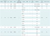
The design of this retrospective study was approved by the Institutional Review Board (Seoul National University Bundang Hospital IRB). From Aug 2011 to Jan 2015, there were 6 thyroid cancer patients who had undergone total thyroidectomy and had suspected bone metastatic lesions based on whole body iodine scintigraphy (WBIS) or BS. Their mean age was 44.7 ± 9.8 years, there was 1 male and 5 female, and 2 cases were papillary thyroid cancer and 4 were follicular thyroid cancer (FTC). Bone PET/CT was then utilized to evaluate the suspected bone lesions. Of the 6 patients, 4 underwent tissue confirmation by computed tomography (CT)-guided needle biopsy (n = 3) or craniectomy (n = 1), and the remaining 2 were clinically followed-up by other methods, such as F-18 fluorodeoxyglucose (FDG) PET/CT, MRI, and serum TG. Characteristics of the patients and lesions were shown in Table 1.
WBIS
Whole body iodine scintigraphy was performed 5–6 days after oral ingestion of I-131 (dose = 5550–7400 MBq). All patients had discontinued long-acting thyroid hormone (T4) replacement 4 weeks prior, and short-acting thyroid hormone (T3) treatment 2 weeks prior to I-131 administration. High iodine intake was prohibited for 2 weeks prior to I-131 administration. A large-field-of-view gamma camera (Discovery NM/CT 670, GE Healthcare, Milwaukee, WI, USA) equipped with a high energy parallel hole collimator was used for imaging.
BS
Bone scintigraphy was performed using a standard protocol. Briefly, 1295 MBq of technetium-99m-hydroxymethylenediphosphonate (99mTc-HDP) (Mallinckrodt, St. Louis, MO, USA) was injected intravenously, and 3 hours thereafter whole body BS images were acquired using a dual-head gamma camera (Forte, ADAC-Philips, Holt, MO, USA) equipped with a low energy high resolution collimator.
Bone PET/CT
Bone PET/CT was performed as described previously (914). Briefly, F-18 sodium fluoride (5.18 MBq/kg body weight) was injected into patients intravenously. Bone PET/CT images were obtained from the skull base to the upper thigh or from the skull vertex to the feet, 30 minutes after the injection. A dedicated PET/CT scanner (DVCT, GE Healthcare, Milwaukee, WI, USA) was used for acquisition. PET images were attenuation-corrected using the CT attenuation map and reconstructed using a 3-dimensional ordered-subset iteration algorithm (VUE point, GE Healthcare, Milwaukee, WI, USA).
Image Interpretation and Determining Malignancy
Whole body iodine scintigraphy, BS, and bone PET/CT images were reviewed by two nuclear medicine physicians, and the final results were based on a consensus between the two experts. WBIS was considered positive for bone metastasis when there was an abnormal lesion with high I-131 uptake over the skeletal area not overlapping with thyroid remnant activity or physiologic uptake in salivary glands, the stomach, or the bladder. Positive findings on BS or bone PET/CT were defined as a skeletal lesion with abnormally increased uptake that differed from a joint or degenerative bony change. Pathologic tissue diagnosis was performed by CT-guided biopsy for 3 lesions of 3 patients, and by craniectomy for 1 skull lesion of 1 patient.
Statistical Analysis
The sensitivity, the specificity, and the accuracy of BS and bone PET/CT for detecting bone metastasis were compared using McNemar's test. Statistical software (MedCalc, version 12.4.0.0, Mariakerke, Belgium) was used for the analyses. P values of < 0.05 were considered statistically significant.
RESULTS
Diagnostic Performances of Bone PET/CT and BS
Seventeen suspected bone metastatic lesions from 6 patients were analyzed. Ten lesions from 2 patients were confirmed bone metastases; whereas, 7 lesions from 4 patients were benign lesions (Table 1). The sensitivity of BS of 20.0% (2/10) was significantly lower than that of bone PET/CT, which was 100% (10/10) (p = 0.008, McNemar's test). The specificities of BS (71.4%, 5/7) and bone PET/CT (57.1%, 4/7) did not differ significantly (p = 1.000, McNemar's test). The accuracy of BS (41.2%, 7/17) was significantly inferior to that of bone PET/CT (82.4%, 14/17; p < 0.025, McNemar's test).
Representative Cases
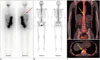
A 54-year-old female patient with FTC undertook second I-131 therapy (5550 MBq), and post-therapy WBIS showed abnormal uptake in the lower T spine area, suggesting metastasis (Fig. 1A). However, there was no definite increase in uptake in the lower T spine area on BS (Fig. 1B). Because TG had been increasing continuously, we decided to perform F-18 FDG PET/CT. However, F-18 FDG PET/CT demonstrated no uptake in the lower T spine area (data not shown). Bone PET/CT was then performed, and increased F-18 uptake was found in the T10 vertebral body (Fig. 1C). This lesion was biopsied and confirmed as metastatic follicular carcinoma.
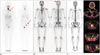
A 42-year-old female patient with FTC had known bone metastasis in the C7 spine. External radiotherapy (39 Gy in total) had been administered over the C7 area before I-131 therapy. In the first post-therapy WBIS (7400 MBq), abnormal uptake was apparent in the right ilium and sternum (Fig. 2A). There was no definite uptake in the right ilium on BS, however, a photopenic defect lesion was apparent in the sternum, and mildly increased uptake was observed in the posterior skull (Fig. 2B). Bone PET/CT was performed to ensure that the right iliac lesion was the metastasis. As a result, it demonstrated multiple areas of uptake in the skull, sternum, C-T-L-S spines, and bilateral ilia suggesting bone metastasis (Fig. 2C). The pathologic diagnosis of the right iliac lesion was metastatic bone lesion originated from thyroid cancer.
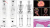
A 53-year-old female patient with FTC and a biopsy-proven bone metastasis in the left proximal femur, underwent wide excision of the metastatic tumor and total thyroidectomy on the same day. I-131 therapy (5550 MBq) was undertaken 3 months later, and post-therapy WBIS showed abnormal uptake in the skull (Fig. 3A). However, BS did not reveal any uptake in the suspected lesion (Fig. 3B). Because the patient had FTC with a proven bone metastasis, bone PET/CT was performed for further clarification. One skull lesion was apparent on the bone PET/CT (Fig. 3C). Based on a suspicion of another bone metastatic lesion in the skull, craniectomy was performed by a neurosurgeon. However, the pathologic diagnosis was a benign inclusion cyst lined with respiratory epithelium (Fig. 3D).
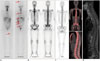
A 50-year-old female FTC patient underwent total thyroidectomy and I-131 therapy (5550 MBq). WBIS showed multiple suspected bone metastatic lesions in the skull, thoracolumbar spines, and left femur (Fig. 4A). However, BS (Fig. 4B) and bone PET/CT (Fig. 4C) were both negative for bone metastases. Furthermore, contrast-enhanced MRI of the whole spines did not show any bone metastatic lesions (Fig. 4D). The stimulated TG level at the time of I-131 therapy was 6.38 ng/mL (normal range: 0–50 ng/mL) with elevated thyroid-stimulating hormone (TSH) (64.45 µIU/mL, normal range: 0.3–4.0 µIU/mL); however, 8 month later, TG was undetectable with TSH level of < 0.05 µIU/mL. Clinically, there were no symptoms or signs of bone metastasis.
DISCUSSION
Bone PET/CT is a validated nuclear imaging tool for investigating metastatic bone lesions in a variety of malignant diseases (91011). In the case of thyroid cancer, 2 previous studies have investigated bone metastasis detection using bone PET/CT (1213). However, the correlation between F-18 uptake positive lesions and pathology results has not been reported previously. One study reported bone metastasis based on findings of a radioactive iodine positive skeletal lesion detected via WBIS with compatible findings on CT and MRI, without further pathologic confirmation (13). In another study, F-18 uptake itself was regarded as the reference standard for bone metastasis, despite the possibility of false positives arising from bone PET/CT (12). The results of the current study indicate the feasibility of bone PET/CT in a small patient cohort, because for the first time, pathologic diagnosis of F-18 positive lesions was investigated in conjunction with BS and WBIS findings.
In the current study, the most striking findings were the benign bone lesions with high I-131 and F-18 uptake (Fig. 3, Table 1). Radioiodine uptake in non-thyroid cancer lesions has been reported in a large number of cases, including a bronchogenic cyst and a parotid gland duct ectasia that accumulated fluids possibly via the sodium iodide symporter, the major gatekeeper of iodine uptake in thyroid cells (151617). On the other hand, F-18 uptake has been reported in diverse benign bone diseases (9). However, the combination of I-131 and F-18 uptake in a benign skeletal lesion of thyroid cancer patient has never been reported in the literature, and requires extra caution during the investigation of thyroid cancer bone metastasis.
In the current study, the superiority of bone PET/CT over planar BS for the detection of bone metastasis was reconfirmed (Figs. 1, 2). The higher sensitivity of bone PET/CT than BS, without the expense of specificity, is concordant with previous studies (910). Single photon emission computed tomography (SPECT) acquisition could increase the diagnostic accuracy of planar BS (13). However, this was beyond the scope of the current study. In a patient, multiple bone lesions suspected on WBIS were not apparent via BS and bone PET/CT (Fig. 4), and all follow-up data including MRI and serum TG levels were negative. The exact nature of the I-131 avid lesions was not clearly addressed at the time of writing this paper. The diagnosis could have been facilitated if I-131 SPECT/CT was performed at the time of I-131 administration or F-18 FDG PET/CT was conducted in follow-up to I-131 treatment. However, bone PET/CT showed the equivalency or at least non-inferiority regarding the exclusion of bone metastasis. Thus, given the importance of detecting metastatic bone lesions in thyroid cancer patients, bone PET/CT may be recommended as the first line imaging modality for the detection/exclusion of bone metastasis, instead of BS.
Limitation of the current study was that F-18 bone PET/CT was not compared with I-131 SPECT/CT and F-18 FDG PET/CT. In thyroid cancer patients with single challenge of I-131 administration, I-131 SPECT/CT was superior to WBIS and F-18 FDG PET/CT, while in patients with multiple challenge of I-131 therapy, F-18 FDG PET/CT was greater than WBIS and I-131 SPECT/CT for detection of bone metastasis (18). Therefore, further studies are required to determine the best imaging modality for detection of thyroid cancer bone
metastasis.
In conclusions, bone PET/CT may be a more useful tool for the diagnosis of bone metastasis of thyroid cancer than BS. However, the possibility of false positives arising from bone PET/CT should be considered, because some benign bone lesions can take up both I-131 and F-18.
 XML Download