

 PDF
PDF ePub
ePub Citation
 Print
Print


INTRODUCTION
Chlorfenapyr is classified as a moderately hazardous pesticide, which has been widely used in eradication of insects over the past 15 years (1). However, chlorfenapyr toxicity in humans has rarely been studied. To date, only 3 cases of human chlorfenapyr intoxication with central nervous system involvement based on radiologic evidence have been reported, all of which were fatal (234). Herein, we reported the first non-fatal case of chlorfenapyr-induced toxic leukoencephalopathy with radiologic reversibility. This report was approved by the Institutional Review Board of our institution and informed consent was waived.
CASE REPORT
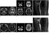
A 44-year-old female patient presented with a 5-day history of bilateral lower leg weakness and urinary incontinence. She had an alert mental status and stable vital signs. A neurologic examination revealed motor weakness in both legs (grade II/grade II) and hypoesthesia below the T11 dermatome. Serum and cerebrospinal fluid laboratory findings were normal. Magnetic resonance imaging (MRI) of the brain and spine was performed using a 1.5T unit (Signa HDxt; GE Medical Systems, Milwaukee, WI, USA). The brain MRI scan revealed extensive and symmetric signal-intensity abnormalities across the entire bilateral cerebral and cerebellar white matter, including the internal capsule, optic nerve and chiasm, corpus callosum, corticospinal tract, and brain stem (Fig. 1). The lesion showed increased signal intensity on T2-weighted images (T2WI) (Fig. 1A), and significant diffusion restriction on diffusion-weighted imaging (DWI) and an apparent diffusion coefficient (ADC) map (Fig. 1B). The spinal MRI scan revealed diffuse swelling and hyperintensity in the entire spinal cord on T2WI (Fig. 1C). The lesion showed no contrast enhancement after intravenous injection of gadolinium (not shown). Based on these MRI findings, toxic leukoencephalopathy was suspected. Detailed patient history revealed that the patient had accidentally ingested a 10% chlorfenapyr solution 19 days prior (2 weeks before symptom onset). The patient had retained the liquid chlorfenapyr orally and immediately spat it out without swallowing. Steroid pulse therapy was initiated as adjunctive treatment for symptom relief. Despite the treatment, neurologic symptoms progressed over the next 7 days. Lower leg weakness progressed to paralysis, and hypoesthesia deteriorated to complete sensory loss below the T8 dermatome. Follow-up MRI performed 71 days later revealed complete resolution of abnormal signal intensities in the brain, brain stem (Fig. 1D, E), and cervical to upper thoracic spinal cord, with residual hyperintensity and atrophic change in the spinal cord below the T7 level (Fig. 1F). During this period, the patient's symptom of paraplegia were unimproved. No other newly developed neurologic deficits were noted. Based on the radiologic and clinical findings, the case was categorized as partially reversible chlorfenapyr-induced toxic leukoencephalopathy.
DISCUSSION
Chlorfenapyr is a widely used pyrrole insecticide (1). Oxidative removal of the N-ethoxymethyl group of chlorfenapyr, using mixed function oxidases, forms the CL 303268 compound. CL 303268 uncouples oxidative phosphorylation at the mitochondria, resulting in disrupted adenosine triphosphate production, cellular death, and mortality of the organism (5). Despite its wide use, there are few reports of chlorfenapyr intoxication in humans (2346).
Radiologic Finding
Based on radiologic evidence, only 3 human chlorfenapyr intoxication cases with central nervous system involvement have been reported (234). Kwon et al. (2) were the first to describe MRI findings of chlorfenapyr-induced leukoencephalopathy. In their report, brain MRI revealed a bilateral, symmetric lesion along the entire white matter tract with high signal intensity on a fluid attenuated inversion recovery scan and diffusion restriction on DWI and an ADC map. The following year, Tharaknath et al. (3) reported another chlorfenapyr intoxication case. MRI scans included in their report revealed diffuse swelling and increased signal intensity in the spinal cord on T2WI, as well as white matter changes in the brain and brain stem, similar to the present case. Recently, Kang et al. (4) also reported a similar case with diffuse, low white matter density on a computed tomography scan. In the present case, brain and spinal MRI showed notably extensive and symmetric hyperintense changes on T2WI confined to the white matter throughout the brain, brain stem, and spine, with significant diffusion restriction. These results are very similar to those of previous cases (234).
In 1998, Kramer et al. reported interesting results based on neurohistological examinations of one-year chlorfenapyr dietary neurotoxicity in rats (1). In their study, a myelinopathic process consisting of vacuolar myelinopathy and mild myelin sheath swelling was found in rats with chlorfenapyr intoxication at 52 weeks. MRI findings, including white matter distribution and high signal intensity on T2WI, reflected these pathologic findings. The reason for pathology of the white matter tracts alone is still unknown. Characteristic involvement of the entire white matter tract in chlorfenapyr intoxication may help distinguish it from other toxic leukoencephalopathy causes such as heroin toxicity or chemotherapeutic agents-induced leukoencephalopathy (7).
Serial MRI findings related to chlorfenapyr-induced leukoencephalopathy have not been previously published. The present case revealed the complete resolution of abnormal signal intensities in the brain and the cervical to upper thoracic spinal cord, with residual hyperintensity and atrophic change in the spinal cord below T7 on follow-up MRI scans obtained 71 days later. There are several reports of diffusion restriction reversibility in follow-up MRI studies assessing toxic leukoencephalopathy with various causes (89). Sivasubramanian et al. (9) referred to this disease entity as "acute reversible toxic leukoencephalopathy". These leukoencephalopathies are being increasingly reported with the increased use of chemotherapeutic agents. However, reversibility of chlorfenapyr-induced leukoencephalopathy has not been reported previously. The pathophysiologic mechanisms of reversible diffusion restriction in toxic leukoencephalopathy are unknown but presumably due to intramyelinic edema, myelin vacuolation, and direct toxic demyelination (8). Similarities between presumed pathomechanisms of radiologic reversibility and pathologic results from animal studies with chlorfenapyr intoxication could explain the radiologic findings observed in the present case.
Clinical Findings
The majority of previous chlorfenapyr intoxication cases exhibited a distinct clinical course that was described as an initial latent period lasting from 7 to 14 days followed by rapidly progressive symptoms resulting in death (2346). Kang et al. (4) assumed that a latent chlorfenapyr intoxication period is related to time required for formation of the active toxic product from the pro-insecticide. A latent period was also observed in our case. The present patient had no symptoms for 14 days after chlorfenapyr ingestion. Because of this latent period, clinicians usually do not consider toxic insults prior to performing an MRI scan.
Whereas most previously reported patients experienced a high fever, diaphoresis, general fatigue, changes in mental state, and death, the present patient only experienced paraplegia despite extensive white matter tract signals. However, previous cases reported an ingested volume of approximately 20 to 250 mL; whereas, the volume ingested by the present patient was estimated to be considerably less, which may have contributed to the non-fatal nature of the neurotoxicity. Ku et al. (10) reported another case of survival after chlorfenapyr intoxication. In this case, the dose consumed was only 10 mL, and the patient experienced mild rhabdomyolysis, fever, and acute pancreatitis. These results suggested that the fatal clinical course from chlorfenapyr intoxication is associated with the ingested volume. Nevertheless, despite the small volume ingested by the patient in the present case, chlorfenapyr ingestion still led to paraplegia. During the follow-up period (71 days), the patient's symptom of paraplegia were unimproved and there were no other persisting or newly developed neurologic deficits on neurologic examination. These clinical findings were consistent with the radiologic findings of partial resolution of abnormal signal intensity with residual hyperintensity and atrophic changes in the thoracic spinal cord.
To our knowledge, this was the first reported case of chlorfenapyr-induced reversible toxic leukoencephalopathy. Serial MRI scans showed resolution of extensive abnormal signal intensities in white matter tracts throughout the brain, brain stem, and spinal cord, with residual hyperintensity and atrophic changes in the lower spinal cord. Unusual radiologic reversibility in the present case was likely related to the small volume of ingested chlorfenapyr. Considering the delayed chlorfenapyr toxicity, MRI features are potentially valuable for prompt diagnosis and adequate follow up in chlorfenapyr-induced toxic leukoencephalopathy cases.
 XML Download