

 PDF
PDF ePub
ePub Citation
 Print
Print


INTRODUCTION
Primary acquired nasolacrimal duct obstruction (PANDO) usually affects women older than 40 years and presents with epiphora. Although its etiology is unclear, many predisposing factors such as previous conjunctival infections, nasal diseases, sinusitis, topical timolol or chloramid exposure are thought to be predisposing factors (123). Some authors consider morphology of bony nasolacrimal duct (BNLD) as a causative factor (456); among these studies, a narrower BNLD transverse diameter (TD) in PANDO patients than the control group is reported (4). In contrast, other studies reported no significant association of BNLD dimensions and PANDO (78). These controversial results make it difficult to reach a definitive conclusion.
It is possible to evaluate BNLD with thin slices in different planes by using multidetector computed tomography (CT). Currently, quantitative measurements are available with advanced post-processing applications for evaluation of BNLD morphology (9). In this study, we aimed to determine the morphometric differences of BNLDs using multidetector CT, in unilateral PANDO patients between PANDO and non-PANDO sides, as compared with the control group.
MATERIALS AND METHODS
Patients
Institutional Review Board approval was obtained before the study. Preoperative non-contrast CT scans of unilateral PANDO patients in a Caucasian population admitted to ophthalmology clinic with epiphora between January and December 2014 were evaluated retrospectively. The diagnosis of PANDO in patients who presented to the ophthalmology clinic with epiphora was confirmed by a lack of patency on lacrimal irrigation. Unilateral PANDO patients who consulted the otorhinolaryngology department preoperatively, and the ones in whom the otorhinolaryngologist requested paranasal CT imaging, were included. The control group was selected from among patients admitted to the ear nose throat clinic without any documented epiphora complaint in the hospital patient database records. Bilateral PANDO patients, and subjects with a previous surgery or trauma history were excluded. Thirty-nine unilateral PANDO patients and 36 subjects for control group were included in the study. Each side of unilateral PANDO patients and control group were evaluated separately. BNLDs in unilateral PANDO patients were divided into two groups i.e., PANDO side and non-PANDO side. Right and left side BNLDs of the control group were accepted as the control group. Finally, the study included 39 PANDO side and 39 non-PANDO side and 72 control BNLDs.
Image Acquisition and Data Achievement
A 64-slice CT scanner (Aquillon 64, Toshiba, Otawara, Japan) was used for image acquisition. Images were obtained in axial plane from the frontal sinuses to nasal floor. Continuous non-overlapping sections of paranasal sinus CT scan were obtained with acquisition parameters of 0.5 mm slice thickness, 120 kV, and 200 mAs. The pixel spacing was 0.3 × 0.3 mm. Images were sent to the workstation (Aquarius Intuition edition ver 4.4.6, TeraRecon, Foster City, CA, USA) for assessment. Reformatted images in sagittal and coronal planes were constituted in addition to axial plane with the same resolution characteristics. Images were evaluated with both bone and soft tissue algorithms.
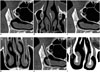
Two radiologists performed measurements separately in the first 20 PANDO patients. Interobserver reliability was determined with intraclass correlation coefficient (ICC); subsequently, one radiologist performed the rest of the measurements in patients and control group. The measurements were as follows: 1) BNLD length (long axis from the proximal end at the lacrimal sac-BNLD junction to the distal end at the level of inferior meatus in sagittal plane) (Fig. 1A) (9), 2) entrance BNLD TD (TD in axial plane at the proximal end of the BNLD) (Fig. 1B) (9), 3) minimum BNLD TD (narrowest TD in axial images) (Fig. 1B), 4) distal end BNLD TD (the TD in axial plane at the distal end of BNLD) (Fig. 1B), 5) BNLD volume (canal volume calculated by Workstation software subsequent to outlining the BNLD wall in all consecutive sagittal images) (Fig. 1C) (8), 6) orientation type (inwards or outwards based on the BNLD orientation with regards to midline in coronal images). Inwards if the orientation is towards the midline and outwards if it is away from the midline (Fig. 1D) (10), 7) orientation angle in sagittal plane (the angle between central line and nasal floor) (Fig. 1E) (5), 8) relative lacrimal sac-BNLD angle (the angle between long axis of lacrimal sac and BNLD in coronal plane) (Fig. 1F) (10).
Statistical Analysis
Shapiro-Wilk test was used to evaluate normality of distribution. Normal distribution was found in all groups for age and BNLD measurements. Therefore, t test was used for age comparison between PANDO patients and control group. One-way ANOVA with post-hoc Tukey test was used for BNLD measurements in PANDO side, non-PANDO side and control groups. Gender difference between patients and controls was evaluated with chi-square test. For the interobserver reliability, ICC was calculated as described by Landis and Koch (11). The male and female measurements were compared within each group separately by Mann-Whitney U test. A p value < 0.05 was considered statistically significant.
RESULTS
There were 9 males and 30 females in the PANDO group and 12 males and 24 females in the control group. Average age was 56.2 ± 14.3 and 59.9 ± 10.2 in PANDO and control groups respectively. Age and gender were not significantly different between patients and control groups (p = 0.200 and 0.320, respectively).
The ICC over 0.820 for each measured parameter indicated excellent agreement between the two observers. In the control group, right and left side measurements were not significantly different; therefore, both eyes were accepted as control eyes (t test, p > 0.300). The mean minimum BNLDs TD in PANDO side and non-PANDO side groups were 3.8 ± 0.8 mm and 3.7 ± 0.8 mm with a range of 2–5.8 mm and 2–5.4 mm, respectively; whereas, in the control group, the mean minimum BNLD TD was 4.1 ± 0.7 mm with a range of 3–6 mm. The distal end BNLD TD was calculated between 2.8–6.3 mm, 2.5–5.8 mm, and 3.6–8.4 mm in PANDO side, non-PANDO side and control groups, respectively. The mean distal end BNLD TDs were 4.6 ± 0.7 mm, 4.5 ± 0.8 mm, and 5.1 ± 0.9 mm, respectively. Although the mean minimum BNLD TD and distal end TD were significantly narrower in both PANDO side and non-PANDO side, as compared to the control group (p < 0.001 and p = 0.040, respectively), there was no difference between PANDO side and non-PANDO side groups. The mean BNLD length, entrance BNLD TD, BNLD volume, orientation angle in sagittal plane and, lacrimal sac-BNLD orientation angle were not statistically different among groups (p > 0.170) (Table 1).
According to BNLD orientation type in coronal plane, the numbers of inward and outward type in the PANDO side group were 25 (64.1%) and 14 (35.9%), respectively; and 24 (61.5%) and 15 (38.5%), respectively, in the non-PANDO side group. Fifty-five (76.4%) inward and 17 (23.6%) outward types were detected in the control group (Table 2). There was no statistically significant difference between PANDO side, non-PANDO side, and control groups (p = 0.190).
In this study, all parameters showed no significant intragroup gender differences in any of the groups (p > 0.050).
DISCUSSION
The BNLD structure is considered as a contributory factor in PANDO development with possible changes in canal morphology causing tear flow resistance (2412). Minimal invasive procedures such as transcanalicular laser therapy, balloon dilatation and stent implantation are in use for diagnosis and treatment of the lacrimal obstructions. Therefore, it is important to know the detailed nasolacrimal duct morphology during management and minimally invasive approaches (13).
CT provides excellent contrast resolution between bony structures and surrounding soft tissues and is therefore, a useful imaging method for BNLD (914). Recently, Takahashi et al. (7) evaluated the BNLD narrowing between the affected and unaffected sides of PANDO patients and control group. They measured the minimum BNLD anteroposterior diameter and minimum BNLD TD. Furthermore, they classified the BNLD shape according to the localization of the minimum BNLD diameter as "funnel" or "hourglass" type. The results of their study indicated a mean minimum BNLD TD of 5.09 ± 1.46 mm, 4.96 ± 1.15 mm, 4.80 ± 0.80 mm in the affected and unaffected sides of PANDO patients and control group, respectively; in addition, there was no difference in mean minimum anteroposterior and TD of BNLD among the groups. Despite the similarity in study design, their results differed from the current study. In their study, the dimensions showed a decreasing trend from PANDO side to non-PANDO side and control group, without significant differences. Janssen et al. (4) compared the mean minimum BNLD TD of PANDO patients who were treated with balloon dacryocystography with control group, but did not consider the non-PANDO side measurements. They reported the mean minimum BNLD TD of 3.5 mm (range, 1.5–6.3 mm) in the control group and 3.0 mm (range 2–4.3 mm) in the patient group, with statistically significant difference. Thus, the smaller BNLD TD was suggested as a predisposing factor for PANDO, since obstruction in the canal might easily develop even with minimal mucosal swelling in the narrowest parts of the canal. This result is consistent with our result and the hypothesis is applicable to ours and previous studies (2615). The BNLD TDs reported by Takahashi et al. (7) were wider in the control and PANDO groups, as compared to those reported by Janssen et al. (4) and the current study and should be considered in interpreting and comparing the results of these three studies. The conflicting result of Takahashi et al. (7) study from the results of Janssen et al. (4) and ours might be related to the effect of ethnicity and race on morphometric measurements.
According to Janssen et al. (4), the minimum mean BNLD TD was significantly narrow in the PANDO patients, however the ranges of diameter found in control group overlapped with the ranges in PANDO patients, suggestive that the narrow BNLD might not be the sole etiologic factor in PANDO development. Likewise, our results showed that narrow BNLD alone could not explain PANDO development.
In our study, the mean minimum BNLD TDs were not statistically different in PANDO side and non-PANDO side groups, as previously reported by Takahashi et al. (7). PANDO has a multifactorial etiology including infections and chronic topical medication use. Therefore, the narrow BNLD may cause a tendency for occlusion of the duct but the environmental factors or exposures, mucus plaques, descending or ascending infections and dacryocystitis attacks may determine the side of the involvement (412).
To the best of our knowledge, the distal end BNLD TD at the level of Hasner's valve was not evaluated in PANDO patients previously. Our results showed that distal end BNLD TD at the level of Hasner's valve was similar in PANDO side and non-PANDO side groups and both were significantly narrower than the control group. The relatively narrow distal end BNLD TD might be a contributory factor that could create a stagnation-like effect in the drainage path of the lacrimal system in PANDO patients that may increase the potential effect of the minimum BNLD TD.
Other parameters calculated in the PANDO patients with multidedector CT were BNLD volume and entrance BNLD TD. Estes et al. (8) recently reported the BNLD volume of the PANDO patients and control group in their study as 0.411 ± 0.18 cm3 and 0.380 ± 0.13 cm3, respectively, without statistical difference. Similarly, there were no significant group-wise differences in our study. Takahashi et al. (7) reported the mean entrance BNLD TD of 6.43 ± 1.38 mm, 6.34 ± 1.37 mm and 6.48 ± 1.30 mm in PANDO side and non-PANDO sides of PANDO patients and control group, respectively; without significant difference among groups, thus corroborating our study results.
The BNLD length was previously reported in the normal population but not in PANDO patients. Ramey et al. (9) reported a BNLD length of 12.3 ± 2.5 mm in men and 10.8 ± 2.5 mm in women in a healthy population. In our study, the mean BNLD length of PANDO patients was similar to the control group and to the healthy population values of Ramey et al. (9). Thus, the length of the canal is not related to the development of PANDO.
It is important to assess the relative lacrimal sac-BNLD angle and BNLD type in coronal plane in PANDO patients to prevent complications before the non-surgical treatment, because inclination between the lacrimal fossa and the entrance of BLND may have variations (16). Relative lacrimal sac-BNLD angle and BNLD type in coronal plane were described in a cadaveric study (10) with mean relative lacrimal sac-BNLD angle of 11.8° with a range of 1–32° and no statistical difference in coronal type of BNLD between genders. In our study, BNLD lengths, coronal and sagittal orientations of BNLD, and relative lacrimal sac-BNLD angles were not statistically different among groups, indicative that they were not causative factors for PANDO.
Primary acquired nasolacrimal duct obstruction is commonly seen in women. Some previous studies reported narrow minimum BNLD TD in women from non-diseased population, and this anatomic characteristic was considered as responsible for PANDO development (5). However, studies performed with PANDO patients show conflicting results. Takahashi et al. (7) revealed narrow BNLD TD in women than men in PANDO side of patients; however, no difference was found in non-PANDO side and control groups. Although Janssen et al. (4) found narrow minimum BNLD TDs in women than men in control group, there was no difference between genders in patient group. This finding is contradictory to the thinking that narrow BNLD in women should be classified as the PANDO side group and not the control group. The same comparison in our study did not reveal any significant difference in terms of gender among PANDO patients and controls. Our results suggest that minimum BNLD TD regardless of gender could be a predisposing factor for PANDO.
Our study revealed significant difference, as compared with control subjects in mean minimum BNLD TD in both PANDO side and non-PANDO side of the PANDO patients. This finding supports the idea that narrow BNLD morphology may cause a tendency for PANDO development. Additionally, narrower distal end BNLD TD detected in both PANDO and non-PANDO sides of unilateral PANDO patients, as compared with control group, may augment the effect of narrow mean minimum BNLD TD. However, lack of difference between PANDO side and non-PANDO side within PANDO patients and some overlap between PANDO patients and control group indicates that the narrow BNLD is not the sole factor.


 XML Download