

 PDF
PDF ePub
ePub Citation
 Print
Print


INTRODUCTION
In recent years, thoracic endovascular aortic repair (TEVAR) has become the most preferred technique for treating thoracic aortic diseases, replacing operative management as the conventional mode of treatment (1). Compared with open surgery, TEVAR is less invasive. Furthermore, TEVAR has better mortality and perioperative outcomes (2345). TEVAR has many advantages, such as single-lung ventilation, aortic cross-clamping, cardiopulmonary bypass, and avoidance of thoracotomy (6). In some patients, however, the passage of an introducer-system into the target site gets impaired by either aortic tortuosity or a lack of mechanical support, which is caused by large aneurysms at the proximal end of the descending thoracic aorta. Therefore, many techniques are used to facilitate stent-graft passage into the target site, such as the use of transbrachial, transseptal, and transapical through-and-through-wire techniques (7891011). In this case report, we have described an alternative technique involving the use of a compliant balloon to provide mechanical support to the proximal descending thoracic aorta, which is required while placing a stent graft into its targeted site. This report was approved by the Institutional Review Board. Moreover, the requirement of an informed consent letter was waived.
CASE REPORT
A 62-year-old man underwent a regular health check-up examination, which detected thoracic aortic aneurysm (TAA). So, he was admitted in the Department of Cardiothoracic Surgery. According to the patient's medical history, he had undergone a car accident 10 years ago and a motor cycle accident 18 years ago. The patient was asymptomatic initially, and a physical examination revealed no significant findings. The patient was on medications for hypertension, which was well under control. The American Society of Anesthesiologists physical status classification system denoted score of one for this patient. The Glasgow Aneurysm Score of this patient was 62 (62 [age in years] + 0 [7 for shock] + 0 [7 for myocardial disease] + 0 [10 for cerebrovascular disease] + 0 [14 for renal disease] = 62). The patient had not undergone any prior vascular surgery. He was hemodynamically stable with a blood pressure of 120/80 mm Hg.
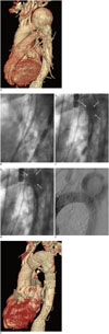
Computed tomography angiography revealed a saccular TAA having a maximum diameter of 63 mm at the greater curvature of the proximal descending thoracic aorta, which was located near the orifice of the left subclavian artery. Therefore, cardiac surgeons decided to perform hybrid TEVAR in zone 2 of this patient. The diameter and length of the proximal neck were 30 mm and 18 mm, respectively. The angulation between the top of the aortic arch and proximal descending thoracic aorta was 81 degrees. The distal neck was 26 mm in diameter without angulation. The radius of the aortic arch was 12 mm. Moreover, the neck of the saccular TAA was wide, having a width of about 5 cm (Fig. 1A). In the CT, we could not detect any development of atheroma, plaque, or thrombus in this patient. So, there were no chances of developing embolism within the aneurysmal sac.
After administering general anesthesia, we first performed bypass surgery on the left common carotid artery and left subclavian artery using an 8-mm vascular prosthesis. This surgery was conducted to maintain arterial flow to the left vertebral and subclavian artery. The proximal left subclavian artery was ligated to prevent type II endoleak. After conducting a successful bypass surgery on the patient, the right common femoral artery was accessed with two sets of Proglide (Proglide; Abbott, Redwood City, CA, USA) using the Preclose-technique. This procedure was performed to enable the entry of the stent-graft's introducer system. To perform control aortography, the left common femoral artery, which was located in the ascending thoracic aorta, was punctured with a 5-Fr marker catheter (Super Torgue; Cordis, Miami Lakes, FL, USA). An extra stiff Lunderquist Guidewire (Cook Medical, Bjaeverskov, Denmark) was inserted through the right common femoral artery sheath into the ascending aorta, over which a 22-Fr introducer system of Zenith Thoracic Pro-Form Endograft (36-157-32 mm; Cook Medical, Bjaeverskov, Denmark) was to be advanced at the target site. However, despite the use of an extra-stiff guidewire, the advancement of the introducer system failed repeatedly, because of a 5-cm wide-necked aneurysm at the greater curvature. A prolapse of the introducer system occurred repeatedly into the wide-necked aneurysm, because the guidewire was not supported by the aortic wall (Fig. 1B).
First, we gently tried transbrachial access through the right subclavian artery. The right subclavian artery was punctured and the snare was inserted; however, this method failed because it was not able to overcome the curvature describing the right subclavian artery, right brachiocephalic artery, and aortic arch.
To facilitate the passage of this introducer system to the target site, a compliant molding balloon (Reliant; Medtronic, Galway, Ireland), having a diameter of, was introduced through a 12-Fr sheath from the contralateral femoral artery. Thus, this balloon was placed within the aneurysm sac. When this compliant balloon was inflated, it created an artificial wall at the greater curvature that was strong enough to support the advancement of the introducer system into the target site. When the operator advanced the introducer system, an assistant pulled the occlusion balloon in inferior direction by carefully providing support to the introducer system. Thus, the assistant prevented an upward prolapse (Fig. 1C). Finally, we were able to successfully advance the introducer system into the target zone (Fig. 1D) and deploy the stent-graft (Fig. 1E). The patient was stable in the peri-operative period, so he was discharged on the sixth day after the operation. After the procedure, the patient did not exhibit any signs of neurological deficits or renal dysfunction. In the 2-year follow-up period, we periodically conducted CT-angiography of the patient. The CT-angiography revealed complete exclusion and shrinkage of the saccular aneurysm along with a patent left subclavian artery bypass graft (Fig. 1F).
DISCUSSION
While performing TEVAR on aortic arch aneurysms, the passage of stent-graft into the intended treatment site is often prohibited due to aortic tortuosity and a lack of mechanical support (7891011). To overcome this problem, researchers have recently recommended the use of wire techniques; these techniques can be used to insert introducer systems into their target zones. We can adopt wire techniques with the following approaches: transbrachial and transseptal access, and snare-assisted pull down of the delivery system (10); the externalized transseptal guidewire technique (78); and the transapical through-and-through-wire technique (911).
In this case, the transbrachial access and through-and-through techniques were first performed gently; however, these techniques failed because we were not able to traverse the curvature formed by the right subclavian artery, right brachiocephalic artery, and aortic arch. This procedure was not performed again as irritation of the neighboring vessels would increase the risk of developing cerebrovascular thromboembolism.
Previous studies have reported that externalized transseptal guidewire techniques provide stability to advancing delivery systems that are introduced in the ascending aorta (10). The advantages of these techniques include enhanced control during stent-graft deployment and conformation of the proximal graft edge (10). However, the externalized guidewire techniques do have some limitations, such as considerable time and effort is expended to create the externalized guide-wire loop. Moreover, an excessive traction may cause injury to an adjacent cardiac structure (7).
Transapical TEVAR can be performed through the left ventricular apex of a beating heart (911). Compared with the traditional retrograde access, the advantages of TEVAR include a short working distance, the ability to advance enough guidewire into the intervention site, and the ability to snare a transapically placed guidewire from a femoral access site (9). However, antegrade transcardiac access techniques may cause injury to the mitral and aortic valves. To repair this damage, additional surgical procedures would have to performed, thereby increasing the operating time.
In this study, we used the balloon-assisted passage technique. This technique has the advantage of utilizing transfemoral access and components readily available in a TEVAR procedure. However, this technique has some disadvantages: it is more prone to over-dilation of balloons, leading to the rupture associated aneurysms. Therefore, we need to inflate the balloon to an appropriate diameter. Furthermore, balloon expansion should be carefully monitored using real-time fluoroscopy. In this case, to avoid drawbacks of this procedure, the balloon was inflated to a diameter that was less than the size of the aneurysm. Thus, we simply created an artificial wall near the aneurysm. In addition, operators must carefully pull the balloon to create a counterforce that would assist the forward movement of stent-grafts, thereby inserting them into the aortic arch. There are many advantages of this technique, including no requirement of further cardiac manipulation and a potentially shorter procedural time.
In summary, the balloon-assisted passage technique reported in this study has facilitated TEVAR in a patient, whose native aortic wall at the greater curvature lacks support to allow the passage of the stent-graft introducer system into the target site. This patient had a wide-necked aneurysm leading to this complication while performing the surgery.
 XML Download