Journal List > Korean J Radiol > v.16(4) > 1027148

Choi, Kim, Kim, Min, Min, Jae, and Chung: Percutaneous Aspiration Embolectomy Using Guiding Catheter for the Superior Mesenteric Artery Embolism

Abstract

Objective

To evaluate the technical feasibility and clinical outcome of percutaneous aspiration embolectomy for embolic occlusion of the superior mesenteric artery (SMA).

Materials and Methods

Between January 2010 and December 2013, 9 patients with embolic occlusion of the SMA were treated by percutaneous aspiration embolectomy in 2 academic teaching hospitals. The aspiration embolectomy procedure was performed with the 6-Fr and 7-Fr guiding catheter. Thrombolysis was performed with urokinase using a multiple-sidehole infusion catheter. The clinical outcome was investigated retrospectively.

Results

Superior mesenteric artery occlusion was initially diagnosed by computed tomography (CT) in all patients, and all patients had no obvious evidence of bowel infarction on CT scan. Percutaneous aspiration embolectomy was primarily performed in 6 patients, and thrombolysis was initially performed in 3 patients. In 3 patients who received primary thrombolysis, percutaneous aspiration was undertaken because the emboli were resistant to urokinase. Complete angiographic success was achieved in 6 patients and partial angiographic success was accomplished in 3 patients. One patient underwent bowel resection. One patient died of whole bowel necrosis and sepsis, and 8 patients survived without complications.

Conclusion

Percutaneous aspiration embolectomy is a useful tool in recanalization of embolic occlusion of the SMA in select patients.

INTRODUCTION

Acute mesenteric ischemia (AMI) comprises 1-2% of acute abdominal emergencies and develops from a sudden decreased perfusion to the intestines (123). Superior mesenteric artery (SMA) embolism is the most common cause of AMI. Although emergent surgical treatment of AMI carries a high morbidity and mortality, the current standard treatment is exploratory laparotomy and surgical removal of the clot (45).
Multidetector computerized tomography (MDCT) has facilitated early diagnosis of SMA occlusion and rapid intervention that improves patient prognosis and decreases the incidence of surgical resection of infarcted bowels. For the past 10 years, many reports have described successfully reperfusion of thromboembolic SMA occlusion by several endovascular strategies such as percutaneous aspiration embolectomy, thrombolysis, percutaneous transluminal angioplasty, primary SMA stenting, and a combination of these therapies, sometimes followed by explorative laparotomy for resection of the infarcted bowel segment (16789101112). However, the case reports or case series deal with patients treated with diverse technique in different institutions. In this study, we described the technical and clinical outcomes of percutaneous aspiration embolectomy in 9 patients with SMA embolism.

MATERIALS AND METHODS

Patients

The Institutional Review Board approved this retrospective study, and informed consent was waived. Between January 2010 and December 2013, 9 patients with orgembolic occlusion of the SMA were treated by percutaneous aspiration embolectomy in 2 academic teaching hospitals. The vascular surgeons and interventional radiologists first attempted endovascular treatment, when MDCT scan had no obvious evidence of bowel infarct.

Interventional Procedures

All percutaneous aspiration procedures were performed by 2 experienced interventional radiologists. All procedures were performed under local anesthesia. The procedure was initiated by inserting a 7-Fr sheath (Super Arrow-Flex PSI set, Arrow International, Reading, PA, USA), 45 cm in length, via the common femoral artery. Selective catheterization of the SMA was performed with the 5-Fr Cobra catheter (C1, Cook, Bloomington, IN, USA). SMA angiography was performed to identify the embolism related filling defect, and then a 7-Fr sheath was engaged at the orifice of the SMA. After a 5-Fr catheter (DAV, Cook) of 110 cm in length was inserted within the 7-Fr guiding catheter (Guider Softip XF, Boston Scientific, Natick, MA, USA) of 90 cm in length, the 7-Fr guiding catheter was introduced into the proximal segment of the SMA. A hydrophilic guidewire (Radifocus, Terumo, Tokyo, Japan) was first navigated into the distal segment of the SMA. Then, a 5-Fr catheter was advanced to the distal segment of SMA over the guidewire and a 7-Fr guiding catheter was advanced over the 5-Fr catheter. After removal of the 5-Fr catheter and guidewire, a 50 mL syringe was connected with the 7-Fr guiding catheter. Aspiration of the emboli was applied manually via the 7-Fr guiding catheter. When performing aspiration, the 7-Fr guiding catheter was removed from the 7-Fr sheath. It was necessary to perform percutaneous aspiration several times to completely remove clots from the main trunk of the SMA. A 6-Fr guiding catheter (Guider Softip XF, Boston Scientific) was used to remove the emboli in the branches of the SMA in the same manner.
Thrombolysis was performed using a multiple-sidehole infusion catheter (Multi-Sideport, Cook) or a microcatheter (Microferret, Cook) in 4 patients. Urokinase (Urokinase-GCC; Green Cross, Seoul, Korea) was infused at a rate of approximately 50000-70000 IU per hour to downsize the emboli or resolve residual emboli.

Analysis

The clinical records, MDCT images and digital subtraction angiography were reviewed retrospectively. Complete angiographic success was pronounced when patency of the SMA and sufficient perfusion of the entire bowel were obtained. If angiography demonstrated residual emboli or sluggish flow of SMA, it was defined as a partial angiographic success. The clinical outcome including laparotomy, bowel resection, hospital stay and mortality was evaluated retrospectively.

RESULTS

Patient Characteristics

The patients included 5 men and 4 women, with an age range of 39-88 years (mean age, 67 years). The clinical characteristics of patients were described in Table 1. SMA occlusion was initially diagnosed by MDCT in all patients. All patients had no obvious evidence of bowel infarct on MDCT scan. SMA embolism was diagnosed by interventional radiologists and vascular surgeon in consensus considering clinical manifestation and radiologic findings.

Endovascular Intervention

Percutaneous aspiration embolectomy was performed in all 9 patients. It was initially undertaken in 6 patients (No. 2-7), and thrombolysis was initially performed in 3 patients (No. 1, 8, 9). Thrombolysis was initially tried in the early study period, and primary aspiration embolectomy was preferentially performed in the late study period. In 5 patients (No. 2, 3, 5-7) primary percutaneous aspiration embolectomy alone, was applied. In 1 patient (No. 4), primary percutaneous aspiration embolectomy was attempted, but residual emboli were noted in the ileal branches. Thrombolysis was conducted for 10 hours, resulting in complete resolution of the emboli. In 3 patients (No. 1, 8, 9) who received primary thrombolysis, percutaneous aspiration embolectomy was undertaken because the emboli were resistant to urokinase. Aspirated emboli consisted of white and red clots (Fig. 1).

Clinical Outcome

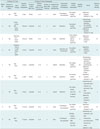
Complete angiographic success was achieved in 6 patients (No. 1, 2, 4-6, 8) and partial angiographic success was achieved in 3 patients (No. 3, 7, 9). Abdominal symptoms resolved within 6 days in the 6 patients with complete angiographic success. Among the 3 patients with partial angiographic success, 1 patient (No. 3) recovered in 2 days without laparotomy. The other patient (No. 7), whose final angiography showed a slow flow in the jejunal branches, underwent laparotomy because she complained of persistent abdominal pain and rebound tenderness was aggravated, resulting in segmental resection of the infarcted small bowel (1.5 meter). In the remaining patient (No. 9), whose final angiography showed multiple residual emboli and a sluggish flow of SMA, whole bowel necrosis was noted during the laparotomy, resulting in death within 3 days. Details of the outcome were listed in Table 2.
Eight patients were discharged without any complication and anticoagulation drugs were prescribed. Two patients (No. 2, 5) had hematochezia after intervention that subsided spontaneously. There was no procedure-related complication such as SMA dissection. Two patients (No. 7, 9) underwent laparotomy and 1 patient (No. 7) received a segmental resection of the small bowel. The 30-day mortality rate was 11% (1 out of 9).

DISCUSSION

A case series of percutaneous SMA aspiration in 15 patients showed 33% (5 of 15 patients) of the 30-day mortality rate (8). In another case series of 10 patients with acute embolic occlusion of the SMA, 1 patient died at 12 hours and another patient died of short bowel syndrome at 8 months (6). Recently, Kawasaki et al. (9) reported 14% (1 of 7 patients) of the 30-day mortality rate and bowel resection in 6 out of 7 patients. In our study, 1 patient needed a segmental bowel resection, and another patient died of sepsis caused by whole bowel necrosis. Although comparison of morbidity and mortality between our study and previous reports has limited value because of small study population, we think that the low morbidity of our study might be explained by the following: 1) patients were diagnosed early by CT scan, 2) patients had relatively mild symptoms and sign, and 3) 3 patients had a partial occlusion of the SMA.
Percutaneous aspiration may be accompanied by the dissection of the SMA and distal embolism. Acosta et al. (6) reported that SMA dissection developed in 2 out of 10 patients with the use of an 8-Fr or 9-Fr guiding catheter. Kawasaki et al. (9) reported 1 dissection out of 7 patients with the 6-Fr guiding catheter. If a larger catheter is used, aspiration of the blood clot is facilitated with the concomitant increased risk of SMA dissection. A guiding catheter may scoop out the intima of an artery during its advancement into the distal SMA, resulting in SMA dissection. Hence, it is best to advance the guiding catheter assembled with a seamless dilator to eliminate the gap between the guidewire and the guiding catheter. Since there is no dilator that fits the guiding catheter, a 5-Fr catheter was used as a dilator. There is no gap between the 6-Fr guiding catheter and the 5-Fr catheter, so the 6-Fr guiding catheter was used to remove the emboli in the branches of the SMA. Although there is a small gap between the 7-Fr guiding catheter and the 5-Fr catheter, careful manipulation of the 7-Fr guiding catheter may prevent SMA dissection.
A distal embolism may develop during advancement of the guiding catheter. A blood clot migrated into the distal arteries can be aspirated by using the 6-Fr guiding catheter or the 5-Fr catheter. If the clot is small and collateral flow is intact, no further treatment is needed because bowel ischemia might not be caused by the migrated clot. If collateral flow is not sufficient, additional thrombolysis can be applied to resolve the migrated blood clot.
Thrombolysis is effective on fresh blood clots, but the emboli that are comprised of old blood clots can be resistant to thrombolysis. In contrast, aspiration embolectomy would likely be effective in the removal of the emboli resistant to thrombolysis. Thus, further study comparing thrombolysis and aspiration embolectomy in the SMA embolism is needed.
In this study, we tried endovascular treatment first when CT scan has no obvious evidence of bowel infarct. Rebound tenderness may suggest transmural bowel necrosis and may lead to exploratory laparotomy in some institutions. However, endovascular treatment may make surgical laparotomy unnecessary or may reduce surgical procedure and time if revascularization of the SMA was achieved even in patients with rebound tenderness. The initial treatment modality should be decided by consensus between interventional radiologist and vascular surgeon considering patient's symptom and sign, CT findings, laboratory results and their clinical experiences. We could achieve good clinical results in select patients with early diagnosis by CT and relatively mild disease severity.
Six patients with complete angiographic success recovered without further treatment such as bowel resection. Of the 3 patients with partial angiographic success, however, 1 patient died and another patient needed segmental bowel resection. Patients with patent SMA and good flow tended to have better clinical outcome in this study.
Several other devices can be used to remove blood clots in the SMA, including a carotid filter, a mechanical rotational thrombectomy device and a MERIC retrieval device (101112). These devices are also effective in the removal of blood clots, but they may not be available in some institutions and may increase medical costs. Aspiration using a guiding catheter is inexpensive and effective according to several studies including the current study (8913).
Transesophageal echocardiography was performed in patient 6 and revealed remnant thrombus in left atrium. In the remaining 6 patients, transthoracic echocardiography was performed and remnant thrombus was not detected in the left atrium. Anticoagulation drugs were prescribed tor all survived patients to prevent re-embolism regardless of findings of echocardiography.
The limitation of our study is the analysis of a small number of enrolled patients that can lead to selection bias and not enough statistical power. Moreover, a comparative study between surgical treatment and endovascular treatment this study was not conducted. Furthermore, overall number of patients with AMI presented at 2 hospitals during the study period is not known.
In conclusion, percutaneous aspiration embolectomy is a useful tool in the recanalization of embolic occlusion of the SMA in select patients.

Figures and Tables

Fig. 1

88-year-old female presented with abdominal pain and hematochezia.
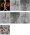
A. Three-dimensional-volume rendered image of abdominal CT angiograph shows segmental occlusion of proximal superior mesenteric artery (SMA) (arrowheads) with calcified plaque. B. Initial angiography shows complete occlusion of SMA (arrowhead) with sluggish flow through distal jejunal branches. Convex meniscus suggests embolic occlusion. C. 7-Fr sheath (arrow) was introduced into proximal segment of SMA, and 7-Fr guiding catheter (arrowhead) was advanced into main trunk of SMA. D. Angiography after aspiration shows partial recanalized SMA and residual blood clot (arrowheads). E. 6-Fr guiding catheter was advanced into distal branch of SMA. F. Final angiography shows complete recanalized SMA. G. Emboli were removed by guiding catheter. Note fresh thrombotic clots (arrowheads) and old embolic clots (arrows).
kjr-16-736-g001
Table 1

Summary of Clinical Features of Nine Patients with SMA Embolism

kjr-16-736-i001
Patient No. Age/Sex Underlying Disease Symptom* Tenderness Rebound Tenderness Laboratory Test* Echocardiography
WBC Count
(× 103/uL)
CRP
(mg/dL)
Serum Creatinine
(mg/dL)
LA Thrombus Regional Wall Motion Abnormality
1 73/M Af, DM Abdominal pain, vomiting, diarrhea Present Present 9.5 15.24 1.16 Absent Absent
2 88/F Af, CAD, CHF Abdominal pain, hematochezia Present Absent 15.5 2.66 1.18 NA
3 72/M Af, hyperthyroidism Abdominal pain, diarrhea, vomitting, dark stool Present Absent 14.2 17.85 0.65 Absent Absent
4 39/F MVR, TVR, pacemaker insertion Abdominal pain Present Absent 10.2 0.59 0.77 Absent Absent
5 61/M CAD, CHF, DM Abdominal pain Present Absent 8.45 0.18 1.37 Absent Severe global hypokinesia
6 55/M CAD, DM Abdominal pain Present Present 7.6 15.2 0.87 Present Inferolateral akinesia
7 66/F Af, DCMP, MR Abdominal pain, vomiting Present Present 16.98 4.68 0.94 NA
8 71/F Pacemaker d/t sick sinus syndrome Abdominal pain Absent Absent 11.4 0.16 1.2 Absent Absent
9 78/M Af, prostate cancer Abdominal pain Present Present 17.6 0.28 1.4 Absent Global hypokinesia of LV

Normal range: WBC count = 4.0-10.0 (× 103/uL), CRP = 0-0.5 (mg/dL), Serum Cr = 0.70-1.40 (mg/dL). *Symptoms and laboratory tests were based on physical examination and blood samples at admission, Transthoracic echocardiography was performed in patients 1, 3, 4, 5, 8, and 9. Transesophageal echocardiography was performed in patient 6. Af = atrial fibrillation, CAD = coronary artery disease, CHF = congestive heart failure, CRP = C-reactive protein, DCMP = dilated cardiomyopathy, DM = diabetes mellitus, LA = left atrium, LV = left ventricle, MR = mitral regurgitation, MVR = mitral valve replacement, NA = not applicable, SMA = superior mesenteric artery, TVR = tricuspid valve replacement, WBC = white blood cell

Table 2

Summary of Intervention and Clinical Outcomes of 9 Patients with SMA Embolism
kjr-16-736-i002
Patient No. Findings of CT Scan Symptom Onset to Angiography Findings of Initial Angiography Intervention (Aspiration, Thrombolysis) Findings of Final Angiography Hospital Stay (d) Result Follow-up Period (Months)
Presence of Hypoperfused Bowel Other Findings Occlusion of Main SMA Trunk Distance between Orifice of SMA and Embolus Number of Patent SMA Branches Collateral Flow to Jejunal/Ileal Branches
1 Yes Mild ileus 3 days Partial 4 cm 5 Good Thrombolysis and aspiration* No residual emboli, good flow 10 Abdominal symptom resolved in 1 day 38
2 Yes Scanty ascites 18 hours Complete 6 cm 3 Slow Aspiration No residual emboli, good flow 9 Hematochezia develop after aspiration, but subsided spontaneously 25
Mild bowel edema Abdominal symptom resolved in 1 day
3 Yes Mild ileus and bowel edema 36 hours Partial 5 cm 8 Good Aspiration Some residual emboli in jejunal branch, good flow 12 Abdominal symptom resolved in 2 days 32
4 No Multifocal infarcts in both kidney and spleen 4 hours Complete 10 cm 7 Good Aspiration and thrombolysis No residual emboli, good flow 11 Abdominal symptom resolved in 4 days 17
5 Yes Mild ileus 4 hours Complete 1 cm 0 Absent Aspiration No residual emboli, good flow 20 Hematochezia develop after aspiration, but subsided spontaneously 5
Abdominal symptom resolved in 6 days
6 Yes Mild ileus 1 week Partial 5 cm 6 Good Aspiration No residual emboli, good flow 22 Abdominal symptom resolved in 1 day 12
7 Yes Mild ileus 5 hours Complete 6 cm 3 Absent Aspiration No residual emboli, slow flow in jejunal branches 16 Laparotomy and segmental bowel resection was performed 1 day after intervention 3
Abdominal symptom resolved in 5 days
8 No Mild ileus 12 hours Complete 8 cm 6 Good Thrombolysis and aspiration No residual emboli, good flow 8 Abdominal symptom resolved in 1 day 32
9 Yes Mild ileus 17 hours Complete 5 cm 2 Slow Thrombolysis and aspiration§ Multiple residual emboli in SMA branches, slow flow 3 Laparotomy was performed 1 day after intervention, and whole bowel necrosis was noted 3 days
Expired in 3 days

*Thrombolysis using urokinase was performed for 24 hours, but no improvement was noted. Then, aspiration thrombectomy was performed, Aspiration thrombectomy was performed, but residual thrombus in ileal branch was noted. Then, thrombolysis using urokinase was performed for 10 hours, Thrombolysis using urokinase was performed for 8 hours, but abundant residual thrombus was noted. Then, aspiration thrombectomy was performed and small residual thrombus in ileal branch was noted. Then additional thrombolysis using urokinase was performed for 6 hours, §Thrombolysis using urokinase was performed for 9 hours, but abundant residual thrombus was noted. Then, aspiration thrombectomy was performed. SMA = superior mesenteric artery

References

1. Björck M, Acosta S, Lindberg F, Troëng T, Bergqvist D. Revascularization of the superior mesenteric artery after acute thromboembolic occlusion. Br J Surg. 2002; 89:923–927.
2. Gupta PK, Natarajan B, Gupta H, Fang X, Fitzgibbons RJ Jr. Morbidity and mortality after bowel resection for acute mesenteric ischemia. Surgery. 2011; 150:779–787.
3. Wong YC, Wu CH, Wang LJ, Chen HW, Lin BC, Huang CC. Mesenteric vascular occlusion: comparison of ancillary CT findings between arterial and venous occlusions and independent CT findings suggesting life-threatening events. Korean J Radiol. 2013; 14:38–44.
4. Park WM, Gloviczki P, Cherry KJ Jr, Hallett JW Jr, Bower TC, Panneton JM, et al. Contemporary management of acute mesenteric ischemia: factors associated with survival. J Vasc Surg. 2002; 35:445–452.
5. Endean ED, Barnes SL, Kwolek CJ, Minion DJ, Schwarcz TH, Mentzer RM Jr. Surgical management of thrombotic acute intestinal ischemia. Ann Surg. 2001; 233:801–808.
6. Acosta S, Sonesson B, Resch T. Endovascular therapeutic approaches for acute superior mesenteric artery occlusion. Cardiovasc Intervent Radiol. 2009; 32:896–905.
7. Resch TA, Acosta S, Sonesson B. Endovascular techniques in acute arterial mesenteric ischemia. Semin Vasc Surg. 2010; 23:29–35.
8. Heiss P, Loewenhardt B, Manke C, Hellinger A, Dietl KH, Schlitt HJ, et al. Primary percutaneous aspiration and thrombolysis for the treatment of acute embolic superior mesenteric artery occlusion. Eur Radiol. 2010; 20:2948–2958.
9. Kawasaki R, Miyamoto N, Oki H, Yamaguchi M, Okada T, Sugimura K, et al. Aspiration therapy for acute superior mesenteric artery embolism with an angled guiding sheath and guiding catheter. J Vasc Interv Radiol. 2014; 25:635–639.
10. Shah SN, Sacks D, Chavali R. Mechanical embolectomy and recanalization of superior mesenteric artery embolism using the MERCI retrieval device. J Vasc Interv Radiol. 2011; 22:1638–1640.
11. Goltz JP, Petritsch B, Spor L, Hahn D, Kickuth R. Acute thromboembolic occlusion of the superior mesenteric artery following covered stent occlusion in the superior mesenteric artery: endovascular therapy using mechanical rotational thrombectomy. Vasa. 2012; 41:375–379.
12. Zeleňák K, Sinák I, Janík J, Mikolajčík A, Mištuna D. Successful recanalization of acute superior mesenteric artery thromboembolic occlusion by a combination of intraarterial thrombolysis and mechanical thrombectomy with a carotid filter. Cardiovasc Intervent Radiol. 2013; 36:844–847.
13. Kim SK, Kwak HS, Chung GH, Han YM. Acute upper limb ischemia due to cardiac origin thromboembolism: the usefulness of percutaneous aspiration thromboembolectomy via a transbrachial approach. Korean J Radiol. 2011; 12:595–601.
TOOLS
Similar articles