INTRODUCTION
Rete mirabile ("wonderful net" in Latin) is a kind of arterial plexus. Carotid rete mirabile (CRM) is one of the collateral patterns formed in the occlusion of the internal carotid artery (ICA). CRM is classically defined to be present at carotid cavernous portion and fed by external carotid artery branches, especially internal maxillary artery (IMA) (1). In spite of its rarity, the rete mirabile has clinical implications in cerebral ischemia or hemorrhage (2, 3). Although the vertebral artery (VA) rete mirabile associated with bilateral CRM has been reported (1), to our knowledge, symptomatic VA stenosis associated with bilateral CRM has not yet been reported. We report the long-term clinical and angiographic outcome after stenting for symptomatic VA stenosis in the patient with bilateral CRM. The Institutional Review Board approved the retrospective review of this case and waived the patient informed consent.
CASE REPORT
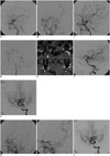
A 22-year-old woman suffered recurrent syncope with increasing frequency, which occurred three times in a week just before admission. All syncope episodes accompanied loss of consciousness for 1 to 2 minutes. She had a mild dull-type headache and nausea for a half day after every episode. She had been healthy before this episode, and no specific diseases were present in her family. Physical and laboratory findings, which included tests for vasculitis, were completely normal. Catheter angiograms revealed a typical appearance of bilateral CRM: bilateral ICA cavernous segments were absent and arterial plexus (rete mirabile) around the cavernous segments reconstituted normal-looking supraclinoid ICA (1). The arterial plexus received their supply from the occluded ICA, IMA, and middle meningeal artery. The ophthalmic arteries (OA) were markedly dilated on both sides and also supplied the supraclinoid ICA by connecting to the rete mirabile. The right VA was occluded, and the left VA showed about 85% stenosis at V4 portion, which gave collateral supply to the bilateral anterior hemisphere through the posterior communicating arteries (PcomA) (Fig. 1A-G). The pre-stenting balloon angioplasty and stent insertion were performed with Voyager RX Coronary Dilatation Catheter 1.5/12 mm and MULTI-LINK VISION Coronary Stent System 4/12 mm (Abbott Vascular, Abbott Park, IL, USA). The post-stenting angiogram showed a significant increase in the collateral supply to the bilateral anterior hemisphere via PcomA. Thereafter, the patient had no recurrence of syncope during the clinical follow-up of 50 months. On the 6-month and the 4-year follow-up angiograms, the bilateral CRM regressed to a faint visualization angiographically, and the stented left VA was entirely responsible for blood supply to the whole brain without any in-stent restenosis (Fig. 1H-J).
DISCUSSION
Angiographic findings generally accepted for the characteristic features of CRM are as follows: 1) hypoplastic ICA; 2) arterial plexus between the maxillary artery and the cavernous portion of the ICA; 3) dilated OA; 4) supraclinoid ICA was not hypoplastic and was fed by the arterial plexus and the OA; 5) bilateral lesions; and 6) no abnormal vessels such as moyamoya vessels in the intradural circulation (1). Our case fully met the angiographic criteria of CRM and also had a severe degree of stenosis of the left VA with contralateral VA occlusion. CRM was considered as a secondary change to ICA regression in the early development or perinatal age when the embryonic arteries were no longer available. Based on this concept and the symmetric feature of CRM, some authors raised the possible presence of a specific trigger or signal to initiate the regression (1, 4). Present but hypoplastic bony carotid canals can be regarded to support this "late regression theory", which is the most accepted one (1, 3, 4). Unfortunately, we could not check the patient's carotid bony canals due to the lack of any brain CT scan, but the source images of the MR angiogram showed probable hypoplastic bony canal (Fig. 1A-G).
It was a deliberating problem whether the lesion of bilateral VA could be a sort of vertebral rete mirabile (VRM). All of the lesions shown in both ICAs and VAs for our case may fall into one category of chronic steno-occlusive disease with prominent collaterals. A few patients with both CRM and VRM have also been reported (1, 4, 5); however, incomplete agreement on the angiographic features of VRM due to its rarity exist. In addition, judging from the fact that most VRM have the segmental absence of their transdural part like CRM (4), VRM could be inferred to have the similar developmental mechanism to CRM. However, which different trigger or mechanism can make a 'concomitant' development of CRM and VRM has not been clarified. In this sense, we cautiously supposed that the lesions of the bilateral VA in our case might be an ongoing process or a specific type of VRM although no proof to support this hypothesis exists. The significance of CRM in humans is not clear. However, its clinical importance is being emphasized by CRM cases presenting with cerebral hemorrhage or ischemia (2, 3).
For cerebral ischemia, research needs to focus on the condition of the collaterals, and so some authors suggested the necessity of proper blood pressure control not too low or some procedures that improve the collaterals (2, 5). In our case, the supraclinoid ICAs were receiving their arterial blood flow not only from CRM but also the posterior circulation through bilateral PcomA. Under this condition, the right VA occlusion and the severe degree of stenosis of left VA likely caused the critical problems. Blood perfusion to both anterior and posterior cerebral circulation became so compromised that it could be a potent cause for the patient's recurrent syncope. In the CRM patient, as commented, it was so important to maintain sufficient collateral circulation that the stenotic left VA should be corrected to improve the whole cerebral perfusion. This inference was quite reasonable when considering that an immediate augmentation of collateral supply to both ICAs via PcomA was possible after stent insertion. In addition, it also strongly supported the inference that the bilateral supraclinoid ICA flow became independent of CRM after stenting and the patient had no recurrence of syncope during the long-term follow-up. As for its developmental mechanism, such post-stent independence might be taken for evidence that the rete mirabile were formed as collaterals to the acquired segmental occlusion but not primarily.
This report is, to our knowledge, the first case of symptomatic VA stenosis associated with bilateral CRM that was treated with stenting. Although most of the bilateral CRM may be asymptomatic and incidentally found, the compromised cerebral circulation can be a significant manifestation of CRM. Therefore, it is important to carefully inspect the patient's collateral circulation because the correction of compromised collaterals can lead an improvement in the patient's clinical condition.


 PDF
PDF ePub
ePub Citation
 Print
Print


 XML Download