

 PDF
PDF ePub
ePub Citation
 Print
Print


INTRODUCTION
Stereotactic radiation therapy was primarily developed for brain neoplasms (1, 2). This therapy applies a high dose of radiation in a defined target volume. Surrounding healthy tissue is not irradiated. This technique has been modified to treat other body tumors (3, 4, 5, 6). The CyberKnife (Accuray, Sunnyvale, CA, USA) system is a recently developed, frameless stereotactic radiosurgery system that tracks tumors in real-time and allows patient movements during the treatment. This system utilizes internal fiducial markers around the target tumor for this purpose.
Stereotactic radiation therapy is also a treatment option for unresectable lung malignancy due to its ability to deliver high doses of conformal radiation to a small target area (3, 4, 5). To precisely target the lung tumors, interventional radiologists are often asked to place markers within the lungs. The standard technique is direct percutaneous transthoracic fiducial placement. The main complications of this method are comprised pneumothorax, migration of fiducial markers and hemorrhage. High rates of pneumothorax (22-67%) with percutaneous transthoracic method have been reported in the literature and might be regarded as an unacceptable risk for some patients with severe cardiopulmonary comorbidity (7, 8). Endovascular placement of a coil as a fiducial marker is a feasible technique in interventional radiology practice and may be a valuable method for such cases. However, only one study has yet been published (7).
The aim of this study was to present our experience with endovascular coil placement for CyberKnife therapy and to describe the technical details and complications of this procedure.
MATERIALS AND METHODS
Patient Demographics
Between June 2005 and September 2013, 163 patients (age range, 35-87 years) with primary or secondary lung malignancies were referred for fiducial placement and subsequent CyberKnife radiosurgery. In 14 patients (9 men, 5 women; mean age, 70 years), direct percutaneous transthoracic fiducial placement was deemed too risky because of a history of pneumonectomy (n = 3), lobectomy (n = 3) or severe cardiopulmonary co-morbidity (n = 8). Indications for radiosurgery were unresectable non-small cell lung carcinoma (n = 11) or metastatic adenocarcinoma (n = 3) in the lungs. The lesions were located in the right upper lobe (n = 4), right lower lobe (n = 2), left upper lobe (n = 5), left lower lobe (n = 3), and mediastinum (n = 1). The largest lesion measured 36 × 41 mm and the smallest lesion was 13 × 10 mm according to World Health Organization criteria (product of largest diameter and greatest perpendicular diameter). The details of these groups are described in Table 1.
Endovascular Coil Placement
Each case was discussed with a radiation oncologist and other responsible primary physicians, and two different interventional radiologists. Medical records were reviewed for previous lung surgery, previous radiation therapy, patient performance status and routine laboratory tests. The benefits and risks of the procedure were fully explained and written informed consent was obtained from each patient.
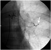
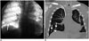
The procedures were performed by interventional radiologists in the biplane angiography suite as a standard pulmonary angiography, which requires percutaneous venous catheterization, intracardiac catheter manipulation and catheterization of the pulmonary artery. A pigtail-type catheter was advanced through the right heart using deflection maneuvers to reach the right or the left pulmonary artery where it was exchanged with a standard 5-Fr diagnostic catheter. A coaxial 2.7-Fr microcatheter (Progreat, Terumo, Tokyo, Japan) was positioned into a distal small pulmonary artery in the vicinity of the lesion, under biplane fluoroscopy guidance. For other lesions not clearly visible on fluoroscopy, previous chest computed tomography (CT) was used to locate the tumor by fluoroscopy in relation to structures like the heart and main vessels. A selective angiogram was done to check the distal vessel size and to confirm the position of the microcatheter, as the maximal distance between coil and tumor should be no more than 50 mm according to the CyberKnife manual. As a final step, a 0.018" 2-mm or 3-mm pushable (Azur, Terumo, Tokyo, Japan) or detachable (Axium, EV3, Irvine, CA, USA and Microplex, Microvention, Columbia, USA) platinum coil was inserted (Fig. 1). In one patient (#5), endovascular coil placement for bilateral lung lesions was done during the same session (Fig. 2A). The patients were discharged on the same day, after a 2-hour post-procedural observation.
Six to nine days after the procedure, noncontrast-enhanced CT was performed for CyberKnife planning before starting radiosurgery. The position and shape of the coils were checked by two interventional radiologists who performed the procedure. Coil-tumor distance, location and coil shape were evaluated on multiplanar CT images (Fig. 2B). Three coils non-collinear in orthogonal planes, with a minimum intercoil distance > 2 cm, maximum coil to tumor distance < 50 mm and a good coiling shape were regarded as successful for this procedure. The numbers, size and tumor-to-coil distance are presented in Table 1.
Late complications of hemorrhage, infarction and infection were also evaluated on CT, along with the clinical status of patients during and after endovascular coil placement to monitor early complications like arrhythmia and vascular complications.
RESULTS
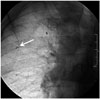
Forty nine metallic micro coils with a median number of 3 coils per tumor were placed with a mean distance of 2.7 cm tumor-to-coil. The planning CT determined that 43 of 49 coils (87.7%) were adequately placed, which were subsequently used as fiducial markers. Two coils could not be used because of larger tumor-coil distance (> 50 mm). Four coils were in acceptable positions, while their non-coiling shapes precluded tumor tracking for the CyberKnife system (Fig. 3). All 14 patients underwent CyberKnife therapy using two or three successfully placed coils, without requiring a repeat endovascular procedure. No long-lasting arrhythmia or vascular complications were noted during catheterization. None of the patients experienced clinical sings of pulmonary complications such as dyspnea, fever and pleuritic pain after endovascular coil placement in early and late phases, as confirmed by CT. No major complications needing further medication other than nominal therapy, overnight hospitalization or permanent adverse sequale were observed.
DISCUSSION
Surgery remains the standard of care for early-stage lung cancer and certain pulmonary metastases (9). In the case of inoperable patients, radiotherapy might be an option. To account for tumor motion in stereotactic radiation therapy planning and treatment delivery, several methods may be used. These include respiratory gating, body frame and real-time tumor tracking. The CyberKnife system utilizes real-time tumor tracking and may require the use of radio-opaque markers implanted near or inside the tumor.
Different methods of marker placement that have been described include percutaneous transthoracic fiducial placement, endovascular coil placement, fiducial placement with bronchoscopy technique and percutaneous extrathoracal fiducial placement (7, 10, 11, 12).
With the standard percutaneous transthoracic puncture, gold seeds or platinum fiducials are placed into the lung. Different size of needles (18 to 25 gauge) from different vendors may be used under guidance of different imaging modalities (ultrasonography, fluoroscopy, CT). Embolization coils may also be implanted by a transthoracic puncture technique. Interestingly, it has been reported that percutaneously implanted platinum endovascular embolization coils are much better seen than standard gold markers, and are successfully tracked by the CyberKnife system (13). However, the percutaneous transthoracic method resulted in a high rate of pneumothorax (22-67%) with tube drainage for the pneumothorax sometimes required. Another common complication is parenchymal hemorrhage, which is usually self-limiting (7, 8). CyberKnife treatment is often performed in elderly patients or patients with co-morbidities like chronic lung disease and previous lung surgery in whom the risk of pneumothorax and parenchymal hemorrhage should be taken seriously. In such patients, endovascular placement of coil as a fiducial marker is a valuable option to avoid these events. We chose the endovascular technique in 14 patients who had symptoms and test results indicating decreased lung reserve due to a previous lung surgery or a chronic lung disease. As in our series, Erasmus Medical Center investigators also found no pneumothorax or parenchymal hemorrhage associated with endovascular delivery of 87 embolization coils in 23 patients (7).
Endovascular fiducial marker placement allowed successful CyberKnife therapy in all 14 patients (100%) in our study, similiar to reports about percutaneous fiducial placement in lung tumors. However, in the percutaneous technique, migration of fiducials in the parenchyma and into the thoracic space is a common problem. This may be evident during the procedure or even in the late phase, necessitating reimplantation (8). During the endovascular procedures, we did not encounter migration of coils, but uncoiling occurred in four patients. Adequacy of coil placement could be determined during the catheterization procedure, which was subsequently confirmed by planning CT.
There are other fiducial implantation techniques, but they might be technically inappropriate for undesired locations and small lung lesions. For example, a small peripheral tumor may be challenging during bronchoscopy, even when using the electromagnetic navigation guidance to reach the tumor (12). Percutaneous placement of fiducial markers into the thoracic wall is another option. However, as in the case of a small central tumor, larger distance and physiological movements precluded tumor tracking for the CyberKnife system. In our experience, the endovascular method is more precise and accurate compared to the other methods of fiducial marker placement, at least in a select group of patients. We believe that the advantage of this technique might be even greater when cone-beam CT is used.
The potential complications of the endovascular procedure in the pulmonary vasculature include vascular injury, thrombosis and pulmonary infarction. Transient pleural pain, hemoptysis and clinically silent small pulmonary infarction have been reported after embolization of pulmonary arteriovenous malformations with different embolic materials as particles, glue and coils (14). The lungs are infrequently infarcted due to dual supply with many anastomoses. The pulmonary and bronchial circulations have two types of connections. The first is an end-to-end anastomosis of 72-325 µm arteries on middle sized bronchi called Sperrarterien. The second type of bronchopulmonary anastomosis is end-to-end or end-to-side, at the precapillary level with a vessel diameter of 24-48 µm (15). The bronchial circulation responds to decreased pulmonary arterial ischemia with dilatation, hypertrophy, and focal proliferation across these meshlike anastomotic channels. In our series, the coils used, unlike liquid embolic material or small particles, probably occluded the vessels mechanically at the proximal sites without inducing widespread thrombosis, which kept these anastomoses open. We did not encounter a patient with a clinical or CT sign of pulmonary infarction. These findings are consistent with other data in the literature (7).
The most common technical problem in our cases was uncoiling and elongation of the coils. We used the smallest coils available on the market. Still we sometimes barely accomplished good coiling shape, given that the smaller diameter in distal vessels close to the lesion could preclude coiling. This problem was more evident with pushable coils, which have no control mechanism to maintain a good coiling shape. Moreover, unlike detachable coils, pushable coils could not be drawn back after insertion as all 4 uncoiled coils in our study. In addition, coils with different chemical properties may have different shape memories and even straining of the coils during manipulations might cause uncoiling.
We did not detect any migration of intravascular coils during follow-up CT scans, probably because they were fixed after placement into a small blood vessel. This seems to be better with hydrocoils which expand after contact with blood. In prior studies, platinum microcoils had enough radio-opacity for the CyberKnife system (7, 13).
In conclusion, endovascular coil placement in distal pulmonary arteries is safe and feasible for CyberKnife radiosurgery. This method might be an option for patients with poor pulmonary condition, in which percutaneous fiducial placement is not safe.

 XML Download