

 PDF
PDF ePub
ePub Citation
 Print
Print


Endoscopic insertion of plastic endoprostheses has become an integral part of the management of many benign and malignant diseases affecting the hepatobiliary and pancreatic system. Clogging and dislocation into the duodenum are the most frequently described complications following stent placement. Distal migration with or without perforation of the colon is an exceedingly rare phenomenon and the treatment is not well defined, as discussed below.
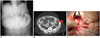
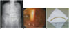
Recently, we were presented with 2 old women with plastic biliary stents (which were endoscopically placed for cholecholithiasis in both cases) that had migrated to their diverticular sigmoid colon, but had different manifestations. The first patient was admitted with the complaint of a relentless pain localized in the lower half of the abdomen: plain film documented stent dislodgement and CT scan revealed that it resided in the sigmoid colon causing perforation (Fig. 1A, B). At laparotomy, the stent was observed to be perforating a diverticulum, and a Hartmann's procedure was accomplished along with stent removal (Fig. 1C). The other patient presented with similar complaints, but the abdomen was more tractable on palpation. Plain abdominal X-ray showed a stent in the lower quadrant of the abdomen and CT scan disclosed a covered perforation of the sigmoid colon (Fig. 2A). Considering the patients' hemodynamically stable condition, colonoscopy was performed with findings of the stent lodged in a diverticular ostium. It was successfully extracted without complications (Fig. 2B, C).
Plastic biliary stent migration to the colon is an extremely rare event: as of 2014, only 25 reports exist in the English literature (1). All these cases dealt with straight plastic stents which were inserted for both benign and malignant diseases of the biliary tree and pancreas (post-cholecystectomy bile leakage and pancreatic adenocarcinoma amongst all) (1). Of these, an uncomplicated endoluminal impaction was observed in 4 cases, whereas perforation occurred in 21 patients. Altogether, the sigmoid colon was the tract most frequently affected (22 cases versus 2 and 1 instance in the right colon and rectum respectively); in particular, among the 21 cases with perforations, 19 accrued from a sigmoid diverticulum (90%), 1 from a non-diverticular sigmoid colon and rectum each. The stiffness and the narrower diameter of the sigmoid tract, together with loci minoris resistentiae, such diverticula represent well-established risk factors for stent anchoring and penetration through the large bowel wall. Currently, there is no standardized diagnostic and therapeutic strategy for this condition (2). Patient clinical status appears to be the main determinant dictating the choice of best management practice. Basically, for endoprostheses that have migrated to the colon without causing perforation, 2 different options are available: the expectant approach and the immediate endoscopic removal. The former is justified by the fact that the vast majority of ingested foreign bodies is commonly expelled without causing any inconvenience, but requires a scrupulous and prolonged examination of the stool, as well as a repeat radiography of the abdomen, whereas the latter is preferred in patients already diagnosed with diverticulosis or inguinal hernia but can itself entail perforation during extraction. All colonic perforation patients were treated surgically in the past several years. However, currently, this treatment is adopted only in the presence of frank peritonitis or fistulous complications (3). On the other hand, in the case of a covered and limited perforation (documented by the proper imaging) and in the absence of peritonism, endoscopic retrieval with clip closure has been proposed as the standard of care (3).
 XML Download