Journal List > Korean J Radiol > v.16(2) > 1027109

Na, Sohn, and Kim: Imaging-Based Management of Acute Ischemic Stroke Patients: Current Neuroradiological Perspectives

Abstract

Advances in imaging-based management of acute ischemic stroke now provide crucial information such as infarct core, ischemic penumbra/degree of collaterals, vessel occlusion, and thrombus that helps in the selection of the best candidates for reperfusion therapy. It also predicts thrombolytic efficacy and benefit or potential hazards from therapy. Thus, radiologists should be familiar with various imaging studies for patients with acute ischemic stroke and the applicability to clinical trials. This helps radiologists to obtain optimal rapid imaging as well as its accurate interpretation. This review is focused on imaging studies for acute ischemic stroke, including their roles in recent clinical trials and some guidelines to optimal interpretation.

INTRODUCTION

Intravenous and endovascular reperfusion therapy are the only proven effective treatment options for acute ischemic stroke patients. Although current advanced stroke imaging has a limited role for time-based intravenous thrombolysis (0-4.5 hours), the role of stroke imaging has expanded substantially to identification of candidates for endovascular therapy and extend the time window of treatment. Optimal stroke imaging management decisions provide crucial information on infarct core, ischemic penumbra/degree of collaterals, vessel occlusion, and thrombus. It is also predictive of benefit or potential hazards (hemorrhage or malignant edema) and thrombolytic efficacy (location of vessel occlusion and extent of thrombus), thereby avoiding futile or unnecessary interventional treatment.
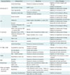
In acute stroke, optimal rapid acquisition and accurate interpretation of imaging studies are of utmost importance to achieve better outcomes. Thus, radiologists should have knowledge on imaging techniques for acute stroke, in addition to interpretation skills. We have reviewed the information on imaging study interpretation for patients with acute stroke and offer a brief summary on the manner and content of imaging reports (Table 1). We also reviewed the current status of imaging-based reperfusion trials (Tables 2, 3, 4).

Understanding Various Imaging Findings and Imaging Strategies in Acute Stroke

Infarct Core Assessment

Unenhanced CT

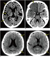
A previous recombinant tissue-type plasminogen activator (rt-PA) trial (1) categorized early ischemic changes (EICs) on baseline unenhanced head CT as follows: 1) focal or diffuse loss of gray/white matter differentiation; 2) focal or diffuse hypodensity or hypoattenuation that is less than the white matter density but greater than cerebrospinal fluid (CSF) density, except for areas of chronic infarcts; 3) focal or diffuse brain swelling with compression of CSF spaces (Fig. 1). Brain swelling, can present with or without concomitant findings of the other 2 categories. Brain swelling without loss of gray/white matter differentiation or hypodense white matter is reportedly not EIC but penumbra (2, 3), hence this so-called isolated cortical swelling is no longer considered EIC. The recent American Heart Association/American Stroke Association (AHA/ASA) guidelines emphasize the implication of "frank hypodensity" on baseline unenhanced CT that affects the treatment scheme using intravenous rt-PA (4). However, there is no clear definition of frank hypodensity in either the guidelines or previous literature. They used "clearly visible mass effect or edema" and considered it as EIC (1). Thus, the second category of EIC mentioned above presumably indicates frank hypodensity; however, the definition of frank hypodensity can be vague and its distinction from loss of gray/white matter differentiation is not explicit on CT.
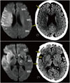
Clinicians can identify whether EIC involves > 1/3 of the middle cerebral artery (MCA) territory (5). However, the extent of EIC may be differently determined among reviewers because EIC > or < 1/3 of the MCA territory is often difficult to determine. The Alberta Stroke Program Early CT (ASPECT) score system devised to improve interrater reliability, is still applied not only to unenhanced CT but also MRI (Fig. 2). However, some issues remain to be resolved: First, there are no anatomic landmarks for distinction of each M region. Second, the interobserver reliability of each region on CT is relatively low (mean intraclass correlation coefficients were 0.640 in M1-M3, 0.530 in M4-M6, 0.762 in the insula, lentiform nucleus, caudate, and 0.367 in the internal capsule) (6).
Information on the ASPECT score that is equivalent to 1/3 the MCA territory is required because some clinicians still prefer the latter. It is difficult to answer this question because the ASPECT score system does not give us an accurate quantified volume of EIC. The presumption is that 1/3 involvement of MCA territory is approximately ASPECT 4-6 (7, 8, 9).
Alberta Stroke Program Early CT 0-4 indicates exclusion of patients from endovascular treatment because of its futility (10). However, some patients with ASPECT < 5 can benefit from endovascular treatment (11). Thus, it is still unknown whether such patients should be excluded or not. If relatively young patients have a lower ASPECT score with salvageable tissue in the eloquent areas, particularly the motor cortex, they may be candidates for mechanical thrombectomy even if they have a higher chance of intracranial hemorrhage (ICH). Thus, we should consider both benefit and risk from treatment when patients have ASPECT < 5.
We should consider both radiation dose and acquisition techniques to obtain optimal unenhanced head CT. The recent guideline from the American College of Radiology describes that the diagnostic reference level and achievable volume CT dose index (CTDIvol) for unenhanced head CT are 75 and 57 mGy, respectively (12). The third CT dose summit recommended the CTDIvol values for each vendor that ranges from 55-60 mGy (13). Helical imaging is faster and can reduce motion artifact compared with sequential imaging. However, it requires a higher radiation dose to obtain imaging quality similar to that of sequential CT at identical imaging parameters because it needs a pitch < 1 (14) and has over ranging. Some recent scanners can minimize over ranging. We can also reduce the radiation dose using noise reduction techniques such as iterative reconstruction. Without considering radiation dose, helical imaging at scanners ≥ 64 detector rows is close to or equivalent to sequential imaging. However, helical imaging at scanners < 16 detector rows tends to have more artifacts (15).
It is important to be aware of the following:
  1. The role of radiologists as an interpreter of baseline unenhanced head CT in acute stroke is to:

    1. Rule out the presence of acute hemorrhage in the brain.

    2. Identify frank hypodensity and report if the extent is > 1/3 of the MCA territory.

    3. Narrow the window width to improve detection of EIC (16), and determine the extent of EIC in the entire brain instead of the 2 planes using ASPECT scores, which is recommended.

  2. The tips for obtaining optimal unenhanced head CT.

    1. Within the recommended CTDIvol for unenhanced head CT (55-60 mGy), we can choose either sequential or helical imaging. The former is superior to the latter in terms of imaging quality at the same imaging parameters, but it is more susceptible to motion-induced artifact. Thus, it is desirable to obtain helical CT when patients are unstable.

    2. It is recommended to use available iterative reconstruction techniques that helps reduce radiation dose while maintaining imaging quality (17).

CT Angiography Source Imaging (CTA-SI)

Although unenhanced CT is the most accessible imaging modality without contraindication, it is sometimes difficult even for experts to identify subtle EIC with relatively less sensitive study. CT angiography source imaging (CTA-SI) is a good alternative and shows higher sensitivity of detection of infarct core than unenhanced CT (18). However, this is the case only when CT angiography (CTA) is obtained with relatively slower scanners. Recent scanners with ≥ 64 detector rows can obtain arterial phase images much faster than old generation scanners, resulting in larger poor contrast-filling areas in cases of major artery occlusion, which may overestimate infarct core (19). CTA-SI, thus, is not a reliable tool to identify infarct core when it is obtained with faster scanners.
It is important to be aware of the following:
CTA-SI is no longer a reliable tool to identify infarct core when it is obtained with faster scanners.

CT Perfusion

Unlike diffusion-weighted imaging (DWI), CT perfusion has caused confusion with regard to the definition of infarct core. At first, an absolute cerebral blood volume (CBV) value of 2.0 mL/100 g was adopted to determine the infarct core (20). Subsequently, it was suggested that a relative cerebral blood flow (rCBF) < 31% threshold best determines infarct core (21). It may be due to different acquisition and/or postprocessing techniques. This issue may remain unresolved until we have a single best technique for CT perfusion.
It is important to be aware of the following:
Absolute CBV or rCBF has been used to determine the infarct core. However, what best represents the infarct core has yet to be determined.

Diffusion-Weighted Imaging (DWI)

Diffusion-weighted imaging is most sensitive and reliable for acute infarct detection. Complete reversal of DWI lesions after reperfusion is limited to tiny lesions in embolic stroke patients (22). Even though reversal post-endovascular reperfusion is attained, it is frequently transient without association with significant salvage of brain tissue or favorable outcomes (23). As such, most lesions with diffusion restriction are generally considered irreversible in clinical practice. However, the exact threshold of ADC value or DWI hyperintensity for irreversibility has not yet been determined.
As in EIC on unenhanced CT, the extent of DWI lesion has high clinical implication (24). Some researchers suggest that patients with DWI lesion > 70 mL (25) or > 100 mL (26) do not benefit from endovascular treatment due to futility. Recent trials adopted the threshold > 70 mL (27) and > 90 mL (28). However, the exact threshold of DWI lesion volume to exclude patients from endovascular treatment has yet to be determined because some patients with larger DWI lesion volumes had favorable outcomes (24, 29). Thus, we cannot entirely rely on the extent of DWI lesion in patient selection for endovascular treatment.
Diffusion-weighted imaging lesion volumes can be easily measured with automated software tools. These tools, however, are not available to all clinicians. DWI ASPECT score can be a good alternative to quantification of DWI lesion volumes. One report suggests that DWI ASPECT < 4 or ≥ 7 may equal to DWI lesion volume > 100 or < 70 mL, respectively (30).
It is important to be aware of the following:
  1. Tips for obtaining better DWI in acute ischemic stroke.

    1. DWI should be obtained at a thickness ≤ 5 mm without a gap.

    2. Thinner DWI often helps in the identification of small acute ischemic lesions in the brainstem.

  2. The role of radiologists as an interpreter of baseline DWI.

    1. Measure DWI lesion volume if software is available, and if not, estimate the volume with the ASPECT score.

    2. DWI may underestimate the acute ischemic lesion, which is more often noted in the basal ganglia (31). Thus, unenhanced CT or fluid-attenuated inversion recovery (FLAIR) images should be evaluated besides DWI.

Assessment of Fluid-Attenuated Inversion Recovery (FLAIR) Imaging

This imaging has recently drawn attention since the finding that a mismatch between DWI and FLAIR is more common in patients who present earlier (Fig. 3). A large retrospective study showed that DWI-FLAIR mismatch identified patients within 4.5 hours of symptom onset with a sensitivity of 62%, specificity of 78%, positive predictive value (PPV) of 83%, and negative predictive value of 54% (32). At a threshold of 3 hours, specificity and PPV of DWI-FLAIR mismatch improved to 93% and 94% (33). However, it still has a shortcoming of relatively lower interobserver reliability (34). Despite this limitation, DWI-FLAIR mismatch is currently under randomized study to determine whether it can improve the outcome in patients of unknown onset with intravenous rt-PA (35).
Hyperintense vessels (HVs) are frequently visualized due to slow flow beyond the occluded site with specificity of 86% and sensitivity of 76% for detection of proximal vascular occlusion (36). They identify proximal occlusion or severe stenosis and may represent the presence of collaterals (Fig. 4). However, robustness of collaterals cannot be assessed by HV alone. Thus, further study to investigate the clinical implication of HV in terms of outcome is required.
It is important to be aware of the following:
  1. DWI-FLAIR mismatch can be used to determine onset time in acute ischemic stroke with a relatively high PPV.

  2. HVs on FLAIR in acute ischemic stroke, represents the presence of proximal occlusion.

Thrombus Assessment

The location of acute thrombus has clinical implication because occlusion in the terminal internal carotid artery (ICA) or basilar artery barely responds to rt-PA (37). Occlusion of such arteries is usually accompanied by a larger thrombus that could explain the lower efficacy of thrombolytic therapy.
The extent of acute thrombus can be determined by using unenhanced CT, CTA, or gradient-recalled echo (GRE) imaging/susceptibility-weighted imaging (SWI). Thin unenhanced CT can detect and measure the length of acute thrombus (38, 39). However, it is not always possible to detect acute thrombus on unenhanced CT. An arterial-phase CTA obtained at faster scanners fails to show contrast filling beyond the occlusion in some patients, which is particularly true in patients with poor collaterals (40). It can be overcome by multiphase imaging such as dynamic CTA or 3-phase CTA that is now adopted for ESCAPE trial (ClinicalTrials.gov NCT01778335). A recent study of dynamic CTA in patients with occlusion in MCA suggested that this technique can predict thrombolytic efficacy thrombus length measurement with a 12 mm cutoff value (41). Another study with unenhanced CT suggested that no thrombus > 8 mm in the MCA is recanalized after intravenous rt-PA (42).
T2* GRE or SWI can also be used to identify acute thrombus in a similar way to that of unenhanced CT (43). However, it is often limited because of the following reasons: First, it may overestimate thrombus extent by dark signal intensity from stagnating blood distal to occlusion. Second, it is prone to artifact, which is problematic at the skullbase. Last, it may not be helpful to characterize thrombus (44).
It is important to be aware of the following:
  1. The location and extent (or length) of thrombus should be determined by unenhanced CT (thinner images increase sensitivity), CTA, or dynamic CTA. Thinner GRE or SWI can approximate the extent of thrombus in the MCA.

  2. Dynamic CTA is the best imaging modality to this end.

Assessment of Hemorrhagic Transformation

Intracranial hemorrhage is a serious complication after intravenous rt-PA treatment. Parenchymal hematoma (PH) can develop in some patients, resulting in poor outcomes. Thus, we need a good imaging biomarker to predict PH prior to treatment. As mentioned earlier, frank hypodensity on unenhanced CT is an important predictor of symptomatic hemorrhage. Larger infarct core may be prone to symptomatic hemorrhage after thrombolytic therapy. However, its sensitivity or specificity is limited because other clinical factors such as higher age, higher stroke severity, and higher glucose are also associated with ICH after rt-PA treatment (45, 46). Assessing damage of the blood-brain barrier can serve as a direct biomarker to predict ICH following thrombolysis, which can be estimated by measuring permeability from CT or MR perfusion (47, 48).
Some researchers suggested that severely reduced CBV (< 2 mL/100 g) on dynamic susceptibility contrast perfusion-weighted imaging (DSC PWI) predicts PH when this area is reperfused after intravenous rt-PA thrombolysis (49).
It is important to be aware of the following:
Extent of infarct core may predict ICH following intravenous rt-PA treatment; however, it is confounded by other clinical factors. CT or MR permeability imaging and very low CBV on DSC PWI have a potential role in this regard.

Imaging Assessment of Cerebral Vascular System

In addition to identification of the presence and location of occlusion, CTA can be used to assess collateral circulation. Collateral circulation is very important because it affects baseline infarct volume, reperfusion, and clinical outcomes (final infarct volume as well) (50). The most reliable assessment tool is conventional angiography. However, not all patients can undergo this invasive procedure. Single-phase CTA has been widely used, but it is limited for accurate assessment of collaterals. Dynamic CTA reconstructed from perfusion CT surmounts this drawback (51, 52). A 3-phase CTA protocol for ESCAPE trial is a good alternative.
CT angiography is usually obtained from the aortic arch to the vertex, which can be helpful for determining the mechanism of stroke and planning for endovascular treatment. It is generally not a requisite before intravenous rt-PA. However, a recent study suggested that pretreatment vascular imaging may help select and stratify patients for trials of thrombolytic therapy (53). Vascular imaging before endovascular treatment is strongly recommended because carotid T- or L-type occlusion or tandem (extracranial or intracranial) ICA and M1 occlusion favors endovascular treatment over intravenous rt-PA (54). Therefore, it would be better to obtain pretreatment CTA in all patients with acute ischemic stroke unless it delays treatment.
Time-of-flight MR angiography (MRA) can also be used to assess occlusion. However, it takes longer to obtain than CTA, and overestimates stenosis and the extent of thrombus. Thus, some clinicians prefer contrast-enhanced MRA (CE MRA) covering the aortic arch up to the intracranial arteries. CE MRA and DSC PWI require separate injection of gadolinium contrast medium that could limit utilization of CE MRA. At 3-T, however, both CE MRA and DSC PWI can be obtained without additional contrast medium by splitting the dose (55).
Susceptibility-weighted imaging can demonstrate prominent asymmetrical cortical and transmedullary veins in the region of ischemia, which possibly represent the region of increased oxygen extraction fraction (Fig. 5). DWI-SWI mismatch may be useful to identify patients who can benefit from reperfusion therapy (56).
It is important to be aware of the following:
  1. Vascular imaging determines the presence and location of occlusion, and is strongly recommended prior to endovascular treatment. It would be beneficial before intravenous rt-PA unless it delays thrombolysis.

  2. Among the noninvasive imaging tools, dynamic or multiphase CTA technique is the best assessment tool for collaterals.

Imaging Assessment of Penumbra

The penumbra can be estimated with CT or MR perfusion imaging and was popular when first introduced. However, a recent randomized trial failed to show its clinical implication (28). There are some issues on CT or MR perfusion: First and foremost, they have not been standardized, which is especially true in CT perfusion (57). While the time to maximum (Tmax) > 6 seconds has recently been chosen to define penumbra on MRI by some researchers (Fig. 6) (27), it is still unclear that this outperforms relative time to peak, mean transit time, or CBF, which are more easily obtained. Second, postprocessing software tools are not standardized, and some of them are commercialized, limiting their availability. A recent study suggests that RAPID (iSchemaview, Stanford, CA, USA) is the best tool (58), but requires further evaluation.
Some agree that assessment of infarct core and collaterals suffices in patient management and expedited endovascular treatment is far more important than penumbra imaging analyses (59). This is supported by the results from IMS III trial (60, 61). Nevertheless, advocates of CT or MR perfusion have enrolled patients in a few clinical trials. Thus, the real value of these advanced imaging will be known in the near future.
It is important to be aware of the following:
  1. CT or MR penumbra imaging has potential for better patient selection and treatment decision.

  2. Rapid assessment of infarct core and collateral circulation and expedited treatment are of utmost importance for attaining better outcomes.

Follow-Up Imaging

Complications, such as hemorrhage after thrombolytic or endovascular treatment are required to be assessed. Differentiation between hemorrhage and contrast enhancement is often difficult on unenhanced CT. Although a recent study suggests that most hyperattenuated lesions following endovascular treatment do not have a significant prognostic value (62), some clinicians still prefer to differentiate them because the fate of hyperattenuated lesions on unenhanced CT obtained immediately after intra-arterial thrombolysis can vary (63), hemorrhage immediately after reperfusion therapy may worsen outcomes, and its growth can be prevented by early discontinuation of antithrombotic medication. Dual-energy CT can be utilized in these cases (64).
Final infarct volume (FIV) used to assess clinical outcomes is determined on imaging obtained at day 30 or 90. It could be alternatively assessed on FLAIR obtained during the first week (days 3-6) (65) or DWI at 24 hours after thrombolysis (66). The importance of 24-hour follow-up imaging is reinforced in a recent study, which claimed that ASPECT score on 24-hour imaging provides better prognostic information compared with baseline ASPECT score (67).
Reperfusion should also be assessed after treatment, because recanalization is not enough to predict final outcomes. Conventional angiography is the best imaging modality, providing angiographic scales such as Modified Thrombolysis in Cerebral Infarction (mTICI) and Thrombolysis in Myocardial Infarction (TIMI). A recent study suggests that mTICI is superior to TIMI in predicting clinical outcome (68). The study shows that an mTICI scale 2b to 3 is optimal to determine procedural success. CT or MR perfusion is another approach for the assessment of reperfusion. A recent study using CT perfusion shows that reperfusion is more strongly associated with good clinical outcome than recanalization (69). Arterial spin labeling MRI can also be used to assess reperfusion (70), which could be useful for patients with poor renal function. Transcranial Doppler ultrasonography would be the best option to monitor reperfusion. However, it is occasionally limited because it cannot penetrate the bony window of all patients, and highly depends on performer skill.
It is important to be aware of the following:
  1. FIV can be estimated with 24-hour follow-up imaging.

  2. Twenty-four-hour imaging also provides better prognostic information than baseline imaging.

  3. Assessment of reperfusion rather than recanalization on 24-hour follow-up CT or MR perfusion helps predict clinical outcomes in patients who do not have endovascular therapy.

Acute Ischemic Stroke Therapy Trials: Current Status and Role of Stroke Imaging

Intravenous Thrombolytic Therapy

Although some are concerned that rt-PA may increase the chance of adverse outcomes through ICH in patients with larger CT EIC, the subsequent analysis of the landmark study (71) showed that the extent of CT EIC does not affect the outcomes after intravenous rt-PA in eligible patients (1). Another retrospective study also shows that intravenous rt-PA should be given to patients within 3 hours of symptom onset, irrespective of the extent of baseline CT EIC although favorable baseline CT (ASPECT > 7) tends to reduce mortality and increase benefit (72). Some clinicians, however, argue that patients with extensive EIC (ASPECT < 3) should not be treated with intravenous rt-PA because of increased risk of ICH (72). The recently published AHA/ASA guidelines suggest CT frank hypodensity > 1/3 of the MCA territory as an exclusion criteria (4). Although this time-based approach is considered very simple and easily applicable, this strategy has a critical weakness because not many patients present within 3 hours after symptom onset. Researchers therefore extend the time limit to treat more patients. Although the first 4 trials of ECASS I (0-6 hours), ECASS II (0-6 hours), ALTANTIS A (0-6 hours), and ATLANTIS B (3-5 hours) could not demonstrate positive results of rt-PA treatment beyond 3 hours (5, 73, 74, 75), a pooled analysis of the previous stroke trials suggest a benefit of rt-PA treatment in the 3-4.5 hour window (76, 77). ECASS III trial proved the benefit of rt-PA and achieved significantly improved outcomes in patients who presented up to 4.5 hours after symptom onset, despite the higher frequency of symptomatic ICH (78). This successful study has led to an official extension of the time limit for intravenous rt-PA up to 4.5 hours, in many countries including South Korea (4).
In ECASS I trial, the CT one-third rule (diffuse swelling of the affected hemisphere, parenchymal hypodensity, and/or effacement of cerebral sulci > 33% of the MCA territory) was introduced for patient selection. Similar CT criteria were used in other stroke trials. Recently, the Third International Stroke Trial recommendation is for CT or MRI only for exclusion of ICH or structural brain lesion mimicking stroke without other CT or MR criteria for patient selection (79). Unlike the previous trials above, EPITHET and DEFUSE studies (3-6-hour time window) adopted advanced MR imaging and showed that intravenous rt-PA significantly attenuates infarct growth and increases reperfusion in most patients with a target mismatch (the presence of PWI/DWI mismatch without a malignant profile) (26, 80, 81). Currently, several clinical trials are evaluating intravenous reperfusion therapy in patients at late time windows (beyond 4.5 hours) (EXTEND, ECASS 4, DIAS 3, and 4) (82, 83, 84) and in those with wake-up stroke by CT or MRI-based selection.

Endovascular Reperfusion Therapy

Although early reperfusion is crucial for the good outcome of reperfusion therapy, the recanalization efficacy of intravenous rt-PA is not as high as endovascular treatment especially when there is occlusion of larger intracranial arteries such as ICA or proximal MCA (85), showing early recanalization rate of 6% and 30% in the terminal ICA and M1, respectively (37). Additionally, a large proportion of patients still present at > 4.5 hours and they are compelled to be excluded from rt-PA therapy. These limitations of rt-PA therapy have prompted the use of endovascular therapy to treat patients contraindicated for rt-PA therapy and to improve recanalization rates. The current guidelines recommend that intra-arterial fibrinolysis is beneficial in carefully selected patients with MCA occlusions within 6 hours of stroke onset (4). The guidelines also permit the use of intra-arterial fibrinolysis or mechanical thrombectomy in patients who have contraindications for rt-PA therapy and in patients with large-artery occlusion who have not responded to intravenous rt-PA therapy (4). There has been a significant increase in the proportion of acute ischemic stroke patients receiving endovascular treatment (86). Advances in endovascular device and technique (87, 88) have facilitated more effective treatment in patients with mechanical thrombectomy when they present within 8 hours of symptom onset. Not all patients, however, benefit from this endovascular treatment. It may be futile, or rather further aggravate. In this context, imaging studies have a pivotal role to select patients who can benefit from endovascular treatment.

Pharmacological Intra-Arterial Thrombolysis

Prolyse in Acute Cerebral Thromboembolism II is the first randomized trial designed to test the safety and effectiveness of intra-arterial recombinant prourokinase (r-pro-UK) to treat MCA (M1 or M2) occlusions within 6 hours of symptom onset (89). Although r-pro-UK-treated group demonstrates an increased recanalization rate and similar mortality compared with the placebo group, r-pro-UK is not US FDA approved. In this trial, CT exclusion criteria included significant mass effect with midline shift and acute hypodense parenchymal lesion or effacement of cerebral sulci in > 1/3 of the MCA territory. Intra-arterial rt-PA thrombolysis or intra-arterial thrombolysis in other locations such as the basilar artery or ICA is based primarily on consensus and case series data.

Mechanical Endovascular Reperfusion Therapy

Mechanical thrombectomy significantly improves recanalization of large artery occlusion compared with pharmacological intra-arterial thrombolysis or clot disruption by a wire manipulation technique. There are currently 4 US FDA approved devices for recanalization that include the earlier MERCI retriever system for distal thrombectomy (90, 91), penumbra aspiration system for proximal thrombectomy (92), recent stent-assisted systems including TREVO (87) and Solitaire (88). The recent trials using stent retrievers (SWIFT and TREVO 2) report higher successful recanalization rates, as compared with the MERCI group (Solitaire 61% vs. MERCI 24%, Trevo 86% vs. MERCI 60%), supporting the superiority of stent-retriever devices to the MERCI device (87, 88).

Recent Randomized Controlled Trials of Intra-Arterial Reperfusion Therapy

Three recent randomized controlled trials (IMS III, SYNTHESIS Expansion, and MR RESCUE) fail to demonstrate any significant benefit of endovascular therapy in acute ischemic stroke (Table 2) (28, 93, 94).
The IMS III trial tested if a combined intravenous rt-PA and intra-arterial endovascular approach is superior to intravenous thrombolysis alone in patients with moderate-to-large ischemic stroke (93). Unfortunately, however, this trial was halted due to futility. In the SYNTHESIS Expansion trial, endovascular therapy for ischemic stroke performed within 4.5 hours of symptom onset was compared with intravenous thrombolysis alone (94). The MR RESCUE trial tested the hypothesis that a favorable CT or MRI penumbral pattern depicted by an automated software program can identify patients likely to achieve greater benefit from endovascular treatment (28).
Table 3 summarizes the major results associated with the outcomes of the 3 endovascular therapy trials. A few points require discussion: First, rapid reperfusion is crucial for good clinical outcome. The subgroup analysis of IMS III trial data demonstrate that there is a significant delay prior to reperfusion, and delays in time to angiographic reperfusion lead to a decreased likelihood of good clinical outcome (60, 61). Although the effect of time delay seems not significant in SYNTHESIS, it might have affected the results of MR RESCUE (28). Second, effective reperfusion depends on mechanical endovascular device. The stent retrieval device which has a higher reperfusion rate than the 1st generation mechanical device is used in only a small number of patients (5%) in the 3 trials (95). Third, the target for endovascular reperfusion therapy should only be patients with large artery occlusion. Only a small portion of patients in IMS III trial and none in SYNTHESIS underwent imaging to determine large artery occlusion leading to selection of patients without large artery occlusion for endovascular therapy. Fourth, imaging-based patient selection is still not established from IMS III and MR RESCUE trials. Although small infarct core (high ASPECT score) and good collateral status strongly predicts good reperfusion and outcome in IMS III trial (11, 96), CT criteria of patient selection for endovascular therapy have yet to be established. The sophisticated multimodal CT or MR model to determine a favorable or unfavorable penumbral pattern fail to identify patients with potential benefit by endovascular treatment in MR RESCUE. Although imaging has the potential to play a key role in selection of optimal patients for endovascular therapy, the best imaging marker requires further investigation. Several ongoing trials of endovascular treatment are designed with advanced CT or MRI in order to select the best candidate for endovascular treatment. The details are described in Table 4.
It is important to be aware of the following:
  1. Intravenous rt-PA should be given to eligible patients with acute ischemic stroke when they present within 4.5 hours after symptom onset.

  2. In patients with frank hypodensity > 1/3 of the MCA territory on unenhanced head CT, intravenous rt-PA should not be given because it is highly associated with subsequent symptomatic ICH.

  3. Intra-arterial fibrinolysis or mechanical thrombectomy can be applied to patients who have contraindications to rt-PA therapy.

  4. Mechanical thrombectomy may be used in patients with large-artery occlusion who have not responded to intravenous rt-PA therapy and may be applied to carefully selected patients who present up to 8 hours after symptom onset. This strategy needs additional randomized trial data and could be changed depending on the results of ongoing trials.

Figures and Tables

Fig. 1

Early ischemic changes on unenhanced head CT (3 different patients).

Unenhanced head CT shows areas of loss of gray/white matter differentiation involving right insula and right temporal lobe (arrows) (A). 83-year-old female with last-seen normal time of approximately midnight underwent CT next day at 8 AM. Attenuation of lesion in right frontal lobe (arrow) is slightly lower than that of contralateral white matter but higher than that of cerebrospinal fluid, suggestive of frank hypodensity (B). Unenhanced CT demonstrates focal gyral swelling with obliteration of adjacent sulci on left (arrows) (C). Note there is no loss of gray/white matter differentiation.
kjr-16-372-g001
Fig. 2

Alberta Stroke Program Early CT (ASPECT) Score.

ASPECT scoring system is applied to both unenhanced CT and diffusion-weighted imaging (DWI). When this system was introduced, it measured scores only at basal ganglia and supraganglionic level. However, it has subsequently evolved to assess entire brain. Normal CT or DWI is scored 10 (3 from subcortical regions and 7 from cortical regions). One point is deducted for each area with abnormality (early ischemic change on CT or lesion showing diffusion restriction). In this particular patient, acute infarct is noted in right M1, M2, M3, M5, I, and L on DWI, yielding ASPECT score of 4. However, it is suggested that right M6 is also affected. This discrepancy may be because ASPECT score does not have landmarks that separate M2 and M3, and M5 and M6. Early ischemic change is also suspected in similar regions on unenhanced head CT (arrows). However, DWI is more sensitive and reliable than unenhanced CT.
kjr-16-372-g002
Fig. 3

Diffusion-weighted imaging (DWI) and fluid-attenuated inversion recovery (FLAIR) mismatch in 76-year-old female.

Last-seen normal time was at 11:00 PM. MRI was obtained on next day at 9:42 AM. Acute infarcts are noted in right middle cerebral artery territory on DWI. However, most DWI lesions do not show hyperintensity in same regions on FLAIR imaging, suggesting that patient had acute infarct within 3 hours.
kjr-16-372-g003
Fig. 4

Hyperintense vessels on fluid-attenuated inversion recovery (FLAIR).

MR angiography shows occlusion in right M1 segment. Hyperintense vessels are noted in branches of right middle cerebral artery on FLAIR images (arrows).
kjr-16-372-g004
Fig. 5

Signs of clot and transmedullary vein involvement on susceptibility-weighted imaging (SWI) in patient with occlusion in right M1 segment.

A. Time-of-flight MR angiography demonstrates occlusion in region of right distal M1 segment. B. Hypointense clot (arrowhead) is noted at corresponding region of right middle cerebral artery on SWI. C. Several hypointense transmedullary veins (arrows) are more conspicuously visualized on right on SWI.
kjr-16-372-g005
Fig. 6

Favorable diffusion-weighted imaging-perfusion-weighted imaging (DWI-PWI) mismatch pattern (large penumbra with small infarct).

A. TOF MR angiography demonstrates occlusion in region of right distal M1 segment. B, C. Lesion on DWI is limited to right insula (B), whereas areas of hypoperfusion (defined by Tmax ≥ 6 seconds [red] and Tmax ≥ 4 seconds [yellow]) are much larger than DWI lesion, representative of favorable DWI-PWI mismatch (C).
kjr-16-372-g006
Table 1

Imaging Studies in Acute Ischemic Stroke: What Should Radiologist Report?

kjr-16-372-i001
Imaging Modality Interpretation Reporting Aims of Imaging
Unenhanced Acute hemorrhage Presence or absence and location Eligibility of further imaging and therapy
Early ischemic change ASPECT score Prediction of outcomes
Eligibility of endovascular therapy
Frank hypodensity ≤ or > 1/3 of MCA territory Eligibility of intravenous rt-PA
Hyperdense artery sign Presence or absence Prediction of thrombolytic efficacy
Location and extent (length)
CTA Acute occlusion Location Prediction of thrombolytic efficacy
Eligibility of endovascular therapy
Collaterals Degree (good, intermediate, or poor) (97) Prediction of reperfusion and outcomes
Eligibility of endovascular therapy
Stenosis ≤ or > 50% Assessment of stroke mechanism
Thrombus (if dynamic CTA available) Length Prediction of thrombolytic efficacy
CT perfusion Infarct core (absolute CBV or relative CBF) Volume Eligibility of endovascular therapy Volume
Prediction of outcomes
Penumbra (Tmax or MTT) Volume, ratio of penumbra to infarct core Eligibility of endovascular therapy
Collaterals (if dynamic CTA available) Degree (excellent, fair, or poor) Prediction of reperfusion and outcomes
DWI Infarct core Volume or ASPECT score Eligibility of endovascular therapy
Prediction of outcomes
Infarct core Location Assessment of stroke mechanism
T2* GRE or SWI Acute hemorrhage Presence or absence and location Eligibility of further imaging and therapy
Susceptibility vessel sign Presence or absence Prediction of thrombolytic efficacy
Location and length
Old microbleeds Number and location Assessment of stroke mechanism
Old hemorrhage Presence or absence and location Assessment of stroke mechanism
FLAIR DWI - FLAIR mismatch Presence or absence and location Eligibility of endovascular therapy
Hyperintense vessel sign Presence or absence and location Determination of occlusion or severe stenosis
MRA Occlusion Presence or absence and location Eligibility of further therapy
Stenosis Presence or absence and location Assessment of stroke mechanism
MR perfusion Penumbra Volume, ratio of penumbra to infarct core Eligibility of endovascular therapy
Follow-up imaging (24 hours after treatment) Infarct core Volume or ASPECT score Prediction of outcomes
Recanalization None, partial, or complete Prediction of outcomes
Reperfusion Index ([baseline lesion volume - follow-up lesion volume] / baseline lesion volume) Prediction of outcomes
Hemorrhagic transformation Presence or absence and location Types (HI 1-2 or PH 1-2) (5) Prediction of outcomes

Note.- ASPECT = Alberta Stroke Program Early CT, CBF = cerebral blood flow, CBV = cerebral blood volume, CTA = CT angiography, DWI = diffusion-weighted imaging, FLAIR = fluid-attenuated inversion recovery, GRE = gradient-recalled echo, HI = hemorrhagic infarction, MCA = middle cerebral artery, MRA = magnetic resonance angiography, MTT = mean transit time, PH = parenchymal hemorrhage, rt-PA = recombinant tissue-type plasminogen activator, SWI = susceptibility-weighted imaging, Tmax = time to maximum

Table 2

Three Randomized Controlled Trials of Endovascular Reperfusion Therapy in Acute Ischemic Stroke Patients

kjr-16-372-i002
Trial Trial Arms Major Clinical Criteria Primary Outcome Primary Results
Safety Efficacy
IMS III 1) IV rt-PA NIHSS score ≥ 10; anterior or posterior circulation; initiation of IV rt-PA within 3 hours of onset; IAT started within 5 hours and completed within 7 hours of onset (time of onset-last time when patient was witnessed to be baseline) mRS score ≤ 2 at 90 days No difference in symptomatic hemorrhage or mortality No difference in good neurological outcome
2) IV rt-PA + endovascular therapy (combined therapy)
SYNTHESIS Expansion 1) IV rt-PA No defined NIHSS threshold; initiation of IV rt-PA within 4.5 hours and IAT within 6 hours from symptom onset mRS score ≤ 2 at 90 days No difference in symptomatic hemorrhage or mortality No difference in good neurological outcome
2) Endovascular
MR RESCUE 1) Embolectomy, penumbral; 2) Standard care, penumbral; 3) Embolectomy, nonpenumbral; 4) Standard care, nonpenumbral; definition of penumbral pattern-infarct core ≤ 90 mL and ratio of volume of penumbral tissue within volume at-risk region (Tmax > 4 s) is > 30% by automated imaging software NIHSS score 6-29; large vessel proximal anterior circulation occlusion; embolectomy can be initiated within 8 hours from symptom onset Shift analysis across 90-day mRS score 0-6 (secondary clinical endpoint - good functional outcome defined as mRS score ≤ 2 at day 90) No difference of 90-day mortality and symptomatic hemorrhage across groups in pairwise comparisons No difference in good outcome (mRS score 0-2) among 4 groups
Embolectomy was not superior to standard care in patients with either favorable penumbral or nonpenumbral pattern

Note.- IMS III = Interventional Management of Stroke III, SYNTHESIS Expansion = Intra-arterial Versus Systemic Thrombolysis for Acute Ischemic Stroke, MR RESCUE = Mechanical Retrieval and Recanalization of Stroke Clots Using Embolectomy. IAT = intra-arterial therapy, IV rt-PA = intravenous recombinant tissue-type plasminogen activator, mRS = modified Rankin Scale, NIHSS = National Institutes of Health Stroke Scale

Table 3

Major Results of Endovascular Reperfusion Therapy

kjr-16-372-i003
Trials Onset Time to Endovascular Therapy Early Reperfusion Rate by Catheter Angiography Endovascular Therapy Method/Device Pretreatment Selection of Large Artery Occlusion Imaging Criteria for Patient Exclusion
IMS III 325 ± 52 minutes (time to termination of procedure)* mTICI 2a-3: 65% (ICA), 81% (M1), 70%/77% (M2 single/multiple occlusion) IA rt-PA (standard or EKOS Microinfusion Catheter System) (most common), various mechanical thrombolysis (no guideline for specific device) Not performed CT: large (more than 1/3 of middle cerebral artery) regions of clear hypodensity on baseline imaging (ASPECTS of ≤ 4 can be used when evaluating > 1/3 MCA). Sulcal effacement and/or loss of grey-white differentiation alone are not contraindications for treatment
mTICI 2b-3: 38% (ICA), 44% (M1), 44%/23% (M2 single/multiple occlusion) If thrombus is not demonstrated, no additional endovascular therapy
SYNTHESIS Expansion Endovascular, 225 minutes; IV rt-PA, 165 minutes (median time to start of treatment) Not provided IA rt-PA (most common) and various mechanical thrombolysis (no guideline for specific device). If no large artery occlusion on angiography, IA rt-PA is still infused Not performed CT: intracranial tumors except small meningiomas, hemorrhage of any degree, severe acute infarction (no specific criteria of extent)
MR RESCUE 381 ± 74 minutes (time to groin puncture) TICI 2a-3, 67% MERCI retriever (most common), penumbra system, IA rt-PA ICA, M1, M2 occlusion by CTA or MRA Proximal ICA occlusion, proximal carotid stenosis > 67% or dissection by contrast-enhanced neck MRA or CTA
TICI 2b-3, 27%

Note.- *From results of IMS III trial data analysis (52). IMS III = Interventional Management of Stroke III, SYNTHESIS Expansion = Intra-arterial Versus Systemic Thrombolysis for Acute Ischemic Stroke, MR RESCUE = Mechanical Retrieval and Recanalization of Stroke Clots Using Embolectomy. CTA = CT angiography, ICA = internal carotid artery, IV rt-PA = intravenous recombinant tissue-type plasminogen activator, MRA = MR angiography, mRS = modified Rankin Scale, mTICI = modified thrombolysis in cerebral infarction, TICI = thrombolysis in cerebral infarction (mTICI 2a grade indicates perfusion of < 1/2 and mTICI 2b indicates perfusion ≥ 1/2 of vascular distribution of occluded artery; TICI 2a grade indicates perfusion < 2/3 and TICI 2b indicates perfusion ≥ 2/3 of vascular distribution of occluded artery)

Table 4

Major Ongoing Imaging-Based Randomized Controlled Trials of Endovascular Reperfusion Therapy Trials

kjr-16-372-i004
Trials Trial Arms Major Clinical Criteria Imaging Modality and Criteria for Patient Inclusion/Exclusion* Endovascular Therapy Method Primary Outcome
MR CLEAN 1) Endovascular therapy 1) NIHSS score ≥ 2 1) Modality: CT or MRI Any method (IA fibrinolysis and any mechanical thrombectomy) mRS score at 90 days
2) Standard care (including IV rt-PA) 2) Possibility to start treatment < 6 hours from onset 2) Inclusion: occlusion of distal ICA or M1/M2 or A1/A2 demonstrated with CTA, MRA, DSA, or TCD
3) Exclusion: not specified
ESCAPE 1) Endovascular therapy 1) NIHSS > 5 at time of randomization 1) Modality: CT Any method (IA fibrinolysis and any mechanical thrombectomy) NIHSS score ≤ 2 or mRS score ≤ 2 at 90 days
2) Standard care (including IV rt-PA) 2) Onset (last seen well) time to randomization time < 12 hours 2) Inclusion: symptomatic intracranial occlusion, on single phase, multiphase or dynamic CTA, at one or more of following locations: Carotid T/L, M1 MCA, or M1-MCA equivalent (2 or more M2-MCAs). Anterior temporal artery is not considered an M2
3) Groin puncture within 60 minutes of CTA 3) Exclusion: unenhanced CT-early ischemic change ASPECT score 0-5, CTA-no or minimal collateral flow > 50%, CTP-low CBV or very low CBF ASPECT score < 6 (if > 8 cm coverage) or > 1/3 MCA (if < 8 cm coverage)
REVACAT 1) Endovascular therapy 1) NIHSS ≥ 6 1) Modality: CT or MRI Solitaire FR Shift analysis across 90-day mRS score 0-6
2) Standard care 2) Treatable (groin puncture) < 8 hours of symptom onset (last seen well) 2) Inclusion: occlusion (TICI 0-1) of the intracranial ICA (distal ICA or T occlusions), MCA-M1 segment or tandem proximal ICA/MCA-M1 suitable for endovascular treatment by CTA, MRA or angiogram, with or without concomitant cervical carotid occlusion or stenosis
3) Ineligible or contraindicated for IV rt-PA, no recanalization after minimum 30 minutes from IV rt-PA 3) Exclusion: ASPECTS < 7 on NCT, CTP-CBV, CTA-SI or ASPECTS < 6 on DWI (diffusion restriction)
POSITIVE 1) Endovascular therapy 1) NIHSS ≥ 8 at time of imaging 1) Modality: CT or MRI Mechanical thrombectomy (aspiration or stent retriever) mRS score at 90 days
2) Standard care 2) Ineligible for IV rt-PA 2) Inclusion: large vessel proximal occlusion (distal ICA through MCA M1 bifurcation)
3) Presenting or persistent symptoms within 12 hours of groin puncture 3) Exclusion: significant mass effect with midline shift or large (> 1/3 MCA) regions of clear hypodensity on NCT or ASPECT score of < 7 (sulcal effacement and/or loss of grey-white differentiation alone are not contraindications for treatment). MR criteria-not provided
THERAPY 1) IV rt-PA + endovascular combined therapy 1) NIHSS criteria: ≥ 8 1) Modality: CT Penumbra system mRS score ≤ 2 at 90 days
2) IV rt-PA 2) Anterior circulation stroke eligible for IV rt-PA 2) Inclusion: large vessel occlusion in anterior circulation with clot length of 8 mm or longer
3) Exclusion: NCT at randomization-significant mass effect with midline shift or large infarct region > 1/3 MCA
EXTEND IA 1) IV rt-PA + endovascular combined therapy 1) NIHSS criteria: not provide 1) Modality: CT or MRI Solitaire FR Reperfusion at 24 hours (CTP or PWI)
2) IV rt-PA 2) Anterior circulation stroke eligible for IV rt-PA within 4.5 hours 2) Inclusion: arterial occlusion on CTA or MRA of the ICA, M1 or M2 + mismatch (Tmax > 6 second delay perfusion volume and CT-rCBF or DWI infarct core volume). Mismatch ratio of greater than 1.2 and absolute mismatch volume > 10 mL NIHSS reduction ≥ 8 or reaching 0-1 at 3 days
3) Treatable (groin puncture) within 6 hours of stroke onset 3) Exclusion: infarct core lesion volume of ≥ 70 mL
SWIFT PRIME 1) IV rt-PA + endovascular combined therapy 1) NIHSS ≥ 8 and < 30 at time of randomization 1) Modality: CT or MRI Solitaire FR mRS score at 90 days
2) IV rt-PA 2) Eligible for IV rt-PA therapy within 4.5 hours of symptom onset (last seen wall) 2) Inclusion: TICI 0-1 flow in terminal ICA, M1 or carotid terminus confirmed by CTA or MRA
3) Treatable < 6 hours of onset of stroke symptoms (last seen well) and < 1.5 hours from CTA or MRA to groin puncture 3) Exclusion: a) hypodensity or MRI hyperintensity > 1/3 of MCA territory (or in other territories, > 100 cc of tissue). b) CT or DWI MRI-moderate/large core defined as extensive early ischemic changes of ASPECT score < 6

Note.- *Exclusion criteria include intracranial hemorrhage on imaging in all trials. MR CLEAN = Multicenter Randomized Clinical Trial of Endovascular Treatment for Acute Ischemic Stroke in the Netherlands, ESCAPE = Endovascular Treatment for Small Core and Proximal Occlusion Ischemic Stroke, REVASCAT = Endovascular Revascularization With Solitaire Device Versus Best Medical Therapy in Anterior Circulation Stroke Within 8 h, POSITIVE = PerfusiOn Imaging Selection of Ischemic STroke PatIents for EndoVascular ThErapy, THERAPY = The Randomized Controlled Trial to Assess the Penumbra System's Safey and Effectiveness in Acute Stroke Treatment, EXTEND IA = Extending the Time for Thrombolysis in Emergency Neurological Deficits-Intra-Arterial, SWIFT PRIME = Solitaire FR as Primary Treatment for Acute Ischemic Stroke. ASPECT = Alberta Stroke Program Early CT, CBF = cerebral blood flow, CBV = cerebral blood volume, CTA = CT angiography, CTP = CT perfusion imaging, DSA = digital subtraction angiography, DWI = diffusion-weighted imaging, FR = flow restoration, IA = intra-arterial, ICA = internal carotid artery, IV rt-PA = intravenous recombinant tissue-type plasminogen activator, MCA = middle cerebral artery, MRA = MR angiography, mRS = modified Rankin Scale, NCT = noncontrast CT, NIHSS = National Institutes of Health Stroke Scale, PWI = perfusion-weighted imaging, TCD = transcranial Doppler, TICI = Thrombolysis in Cerebral Infarction classification

Acknowledgments

We would like to thank Dr. Shang Hun Shin for generously providing figures.

Notes

This work was supported by the National Research Foundation of Korea (NRF) grant (NRF-2013M3A9B2076548).

References

1. Patel SC, Levine SR, Tilley BC, Grotta JC, Lu M, Frankel M, et al. Lack of clinical significance of early ischemic changes on computed tomography in acute stroke. JAMA. 2001; 286:2830–2838.
2. Na DG, Kim EY, Ryoo JW, Lee KH, Roh HG, Kim SS, et al. CT sign of brain swelling without concomitant parenchymal hypoattenuation: comparison with diffusion- and perfusion-weighted MR imaging. Radiology. 2005; 235:992–998.
3. Butcher KS, Lee SB, Parsons MW, Allport L, Fink J, Tress B, et al. Differential prognosis of isolated cortical swelling and hypoattenuation on CT in acute stroke. Stroke. 2007; 38:941–947.
4. Jauch EC, Saver JL, Adams HP Jr, Bruno A, Connors JJ, Demaerschalk BM, et al. Guidelines for the early management of patients with acute ischemic stroke: a guideline for healthcare professionals from the American Heart Association/American Stroke Association. Stroke. 2013; 44:870–947.
5. Hacke W, Kaste M, Fieschi C, Toni D, Lesaffre E, von Kummer R, et al. The European Cooperative Acute Stroke Study (ECASS). Intravenous thrombolysis with recombinant tissue plasminogen activator for acute hemispheric stroke. JAMA. 1995; 274:1017–1025.
6. Finlayson O, John V, Yeung R, Dowlatshahi D, Howard P, Zhang L, et al. Interobserver agreement of ASPECT score distribution for noncontrast CT, CT angiography, and CT perfusion in acute stroke. Stroke. 2013; 44:234–236.
7. Demaerschalk BM, Silver B, Wong E, Merino JG, Tamayo A, Hachinski V. ASPECT scoring to estimate >1/3 middle cerebral artery territory infarction. Can J Neurol Sci. 2006; 33:200–204.
8. Dzialowski I, Hill MD, Coutts SB, Demchuk AM, Kent DM, Wunderlich O, et al. Extent of early ischemic changes on computed tomography (CT) before thrombolysis: prognostic value of the Alberta Stroke Program Early CT Score in ECASS II. Stroke. 2006; 37:973–978.
9. Puetz V, Dzialowski I, Hill MD, Demchuk AM. The Alberta Stroke Program Early CT Score in clinical practice: what have we learned? Int J Stroke. 2009; 4:354–364.
10. Yoo AJ, Zaidat OO, Chaudhry ZA, Berkhemer OA, González RG, Goyal M, et al. Impact of pretreatment noncontrast CT Alberta Stroke Program Early CT Score on clinical outcome after intra-arterial stroke therapy. Stroke. 2014; 45:746–751.
11. Hill MD, Demchuk AM, Goyal M, Jovin TG, Foster LD, Tomsick TA, et al. Alberta Stroke Program early computed tomography score to select patients for endovascular treatment: Interventional Management of Stroke (IMS)-III Trial. Stroke. 2014; 45:444–449.
12. ACR-AAPM. Practice parameter for diagnostic reference levels and achievable doses in medical X-ray imaging. Reston: American College of Radiology;2014.
13. Supanich MP. Protocol Review - Interactive Session: what are the participants using for head CT? Phoenix: AAPM;2013.
14. Bahner ML, Reith W, Zuna I, Engenhart-Cabillic R, van Kaick G. Spiral CT vs incremental CT: is spiral CT superior in imaging of the brain? Eur Radiol. 1998; 8:416–420.
15. AAPM. Adult Routine Head CT Protocols Version 1.1. College Park: American Association of Physicists in Medicine;2012.
16. Lev MH, Farkas J, Gemmete JJ, Hossain ST, Hunter GJ, Koroshetz WJ, et al. Acute stroke: improved nonenhanced CT detection--benefits of soft-copy interpretation by using variable window width and center level settings. Radiology. 1999; 213:150–155.
17. Rapalino O, Kamalian S, Kamalian S, Payabvash S, Souza LC, Zhang D, et al. Cranial CT with adaptive statistical iterative reconstruction: improved image quality with concomitant radiation dose reduction. AJNR Am J Neuroradiol. 2012; 33:609–615.
18. Bhatia R, Bal SS, Shobha N, Menon BK, Tymchuk S, Puetz V, et al. CT angiographic source images predict outcome and final infarct volume better than noncontrast CT in proximal vascular occlusions. Stroke. 2011; 42:1575–1580.
19. Pulli B, Schaefer PW, Hakimelahi R, Chaudhry ZA, Lev MH, Hirsch JA, et al. Acute ischemic stroke: infarct core estimation on CT angiography source images depends on CT angiography protocol. Radiology. 2012; 262:593–604.
20. Wintermark M, Flanders AE, Velthuis B, Meuli R, van Leeuwen M, Goldsher D, et al. Perfusion-CT assessment of infarct core and penumbra: receiver operating characteristic curve analysis in 130 patients suspected of acute hemispheric stroke. Stroke. 2006; 37:979–985.
21. Campbell BC, Christensen S, Levi CR, Desmond PM, Donnan GA, Davis SM, et al. Comparison of computed tomography perfusion and magnetic resonance imaging perfusion-diffusion mismatch in ischemic stroke. Stroke. 2012; 43:2648–2653.
22. Albach FN, Brunecker P, Usnich T, Villringer K, Ebinger M, Fiebach JB, et al. Complete early reversal of diffusion-weighted imaging hyperintensities after ischemic stroke is mainly limited to small embolic lesions. Stroke. 2013; 44:1043–1048.
23. Inoue M, Mlynash M, Christensen S, Wheeler HM, Straka M, Tipirneni A, et al. Early diffusion-weighted imaging reversal after endovascular reperfusion is typically transient in patients imaged 3 to 6 hours after onset. Stroke. 2014; 45:1024–1028.
24. Olivot JM, Mosimann PJ, Labreuche J, Inoue M, Meseguer E, Desilles JP, et al. Impact of diffusion-weighted imaging lesion volume on the success of endovascular reperfusion therapy. Stroke. 2013; 44:2205–2211.
25. Yoo AJ, Verduzco LA, Schaefer PW, Hirsch JA, Rabinov JD, González RG. MRI-based selection for intra-arterial stroke therapy: value of pretreatment diffusion-weighted imaging lesion volume in selecting patients with acute stroke who will benefit from early recanalization. Stroke. 2009; 40:2046–2054.
26. Albers GW, Thijs VN, Wechsler L, Kemp S, Schlaug G, Skalabrin E, et al. Magnetic resonance imaging profiles predict clinical response to early reperfusion: the diffusion and perfusion imaging evaluation for understanding stroke evolution (DEFUSE) study. Ann Neurol. 2006; 60:508–517.
27. Lansberg MG, Straka M, Kemp S, Mlynash M, Wechsler LR, Jovin TG, et al. MRI profile and response to endovascular reperfusion after stroke (DEFUSE 2): a prospective cohort study. Lancet Neurol. 2012; 11:860–867.
28. Kidwell CS, Jahan R, Gornbein J, Alger JR, Nenov V, Ajani Z, et al. A trial of imaging selection and endovascular treatment for ischemic stroke. N Engl J Med. 2013; 368:914–923.
29. Inoue M, Olivot JM, Labreuche J, Mlynash M, Tai W, Albucher JF, et al. Impact of diffusion-weighted imaging Alberta stroke program early computed tomography score on the success of endovascular reperfusion therapy. Stroke. 2014; 45:1992–1998.
30. de Margerie-Mellon C, Turc G, Tisserand M, Naggara O, Calvet D, Legrand L, et al. Can DWI-ASPECTS substitute for lesion volume in acute stroke? Stroke. 2013; 44:3565–3567.
31. Kawano H, Hirano T, Nakajima M, Inatomi Y, Yonehara T. Diffusion-weighted magnetic resonance imaging may underestimate acute ischemic lesions: cautions on neglecting a computed tomography-diffusion-weighted imaging discrepancy. Stroke. 2013; 44:1056–1061.
32. Thomalla G, Cheng B, Ebinger M, Hao Q, Tourdias T, Wu O, et al. DWI-FLAIR mismatch for the identification of patients with acute ischaemic stroke within 4·5 h of symptom onset (PRE-FLAIR): a multicentre observational study. Lancet Neurol. 2011; 10:978–986.
33. Thomalla G, Rossbach P, Rosenkranz M, Siemonsen S, Krützelmann A, Fiehler J, et al. Negative fluid-attenuated inversion recovery imaging identifies acute ischemic stroke at 3 hours or less. Ann Neurol. 2009; 65:724–732.
34. Galinovic I, Puig J, Neeb L, Guibernau J, Kemmling A, Siemonsen S, et al. Visual and region of interest-based inter-rater agreement in the assessment of the diffusion-weighted imaging-fluid-attenuated inversion recovery mismatch. Stroke. 2014; 45:1170–1172.
35. Thomalla G, Fiebach JB, Østergaard L, Pedraza S, Thijs V, Nighoghossian N, et al. A multicenter, randomized, double-blind, placebo-controlled trial to test efficacy and safety of magnetic resonance imaging-based thrombolysis in wake-up stroke (WAKE-UP). Int J Stroke. 2014; 9:829–836.
36. Cheng B, Ebinger M, Kufner A, Köhrmann M, Wu O, Kang DW, et al. Hyperintense vessels on acute stroke fluid-attenuated inversion recovery imaging: associations with clinical and other MRI findings. Stroke. 2012; 43:2957–2961.
37. Bhatia R, Hill MD, Shobha N, Menon B, Bal S, Kochar P, et al. Low rates of acute recanalization with intravenous recombinant tissue plasminogen activator in ischemic stroke: real-world experience and a call for action. Stroke. 2010; 41:2254–2258.
38. Riedel CH, Jensen U, Rohr A, Tietke M, Alfke K, Ulmer S, et al. Assessment of thrombus in acute middle cerebral artery occlusion using thin-slice nonenhanced Computed Tomography reconstructions. Stroke. 2010; 41:1659–1664.
39. Riedel CH, Zoubie J, Ulmer S, Gierthmuehlen J, Jansen O. Thin-slice reconstructions of nonenhanced CT images allow for detection of thrombus in acute stroke. Stroke. 2012; 43:2319–2323.
40. Frölich AM, Schrader D, Klotz E, Schramm R, Wasser K, Knauth M, et al. 4D CT angiography more closely defines intracranial thrombus burden than single-phase CT angiography. AJNR Am J Neuroradiol. 2013; 34:1908–1913.
41. Rohan V, Baxa J, Tupy R, Cerna L, Sevcik P, Friesl M, et al. Length of occlusion predicts recanalization and outcome after intravenous thrombolysis in middle cerebral artery stroke. Stroke. 2014; 45:2010–2017.
42. Riedel CH, Zimmermann P, Jensen-Kondering U, Stingele R, Deuschl G, Jansen O. The importance of size: successful recanalization by intravenous thrombolysis in acute anterior stroke depends on thrombus length. Stroke. 2011; 42:1775–1777.
43. Weisstanner C, Gratz PP, Schroth G, Verma RK, Köchl A, Jung S, et al. Thrombus imaging in acute stroke: correlation of thrombus length on susceptibility-weighted imaging with endovascular reperfusion success. Eur Radiol. 2014; 24:1735–1741.
44. Fujimoto M, Salamon N, Mayor F, Yuki I, Takemoto K, Vinters HV, et al. Characterization of arterial thrombus composition by magnetic resonance imaging in a swine stroke model. Stroke. 2013; 44:1463–1465.
45. Whiteley WN, Slot KB, Fernandes P, Sandercock P, Wardlaw J. Risk factors for intracranial hemorrhage in acute ischemic stroke patients treated with recombinant tissue plasminogen activator: a systematic review and meta-analysis of 55 studies. Stroke. 2012; 43:2904–2909.
46. Strbian D, Engelter S, Michel P, Meretoja A, Sekoranja L, Ahlhelm FJ, et al. Symptomatic intracranial hemorrhage after stroke thrombolysis: the SEDAN score. Ann Neurol. 2012; 71:634–641.
47. Aviv RI, d'Esterre CD, Murphy BD, Hopyan JJ, Buck B, Mallia G, et al. Hemorrhagic transformation of ischemic stroke: prediction with CT perfusion. Radiology. 2009; 250:867–877.
48. Leigh R, Jen SS, Hillis AE, Krakauer JW, Barker PB. STIR and VISTA Imaging Investigators. Pretreatment blood-brain barrier damage and post-treatment intracranial hemorrhage in patients receiving intravenous tissue-type plasminogen activator. Stroke. 2014; 45:2030–2035.
49. Campbell BC, Christensen S, Parsons MW, Churilov L, Desmond PM, Barber PA, et al. Advanced imaging improves prediction of hemorrhage after stroke thrombolysis. Ann Neurol. 2013; 73:510–519.
50. Liebeskind DS. Collateral lessons from recent acute ischemic stroke trials. Neurol Res. 2014; 36:397–402.
51. Frölich AM, Wolff SL, Psychogios MN, Klotz E, Schramm R, Wasser K, et al. Time-resolved assessment of collateral flow using 4D CT angiography in large-vessel occlusion stroke. Eur Radiol. 2014; 24:390–396.
52. Smit EJ, Vonken EJ, van Seeters T, Dankbaar JW, van der Schaaf IC, Kappelle LJ, et al. Timing-invariant imaging of collateral vessels in acute ischemic stroke. Stroke. 2013; 44:2194–2199.
53. González RG, Furie KL, Goldmacher GV, Smith WS, Kamalian S, Payabvash S, et al. Good outcome rate of 35% in IV-tPA-treated patients with computed tomography angiography confirmed severe anterior circulation occlusive stroke. Stroke. 2013; 44:3109–3311.
54. Demchuk AM, Goyal M, Yeatts SD, Carrozzella J, Foster LD, Qazi E, et al. Recanalization and clinical outcome of occlusion sites at baseline CT angiography in the Interventional Management of Stroke III trial. Radiology. 2014; 273:202–210.
55. Nael K, Meshksar A, Ellingson B, Pirastehfar M, Salamon N, Finn P, et al. Combined low-dose contrast-enhanced MR angiography and perfusion for acute ischemic stroke at 3T: a more efficient stroke protocol. AJNR Am J Neuroradiol. 2014; 35:1078–1084.
56. Lou M, Chen Z, Wan J, Hu H, Cai X, Shi Z, et al. Susceptibility-diffusion mismatch predicts thrombolytic outcomes: a retrospective cohort study. AJNR Am J Neuroradiol. 2014; 35:2061–2067.
57. Bivard A, Levi C, Spratt N, Parsons M. Perfusion CT in acute stroke: a comprehensive analysis of infarct and penumbra. Radiology. 2013; 267:543–550.
58. Churilov L, Liu D, Ma H, Christensen S, Nagakane Y, Campbell B, et al. Multiattribute selection of acute stroke imaging software platform for Extending the Time for Thrombolysis in Emergency Neurological Deficits (EXTEND) clinical trial. Int J Stroke. 2013; 8:204–210.
59. Goyal M, Menon BK, Derdeyn CP. Perfusion imaging in acute ischemic stroke: let us improve the science before changing clinical practice. Radiology. 2013; 266:16–21.
60. Goyal M, Almekhlafi MA, Fan L, Menon BK, Demchuk AM, Yeatts SD, et al. Evaluation of interval times from onset to reperfusion in patients undergoing endovascular therapy in the Interventional Management of Stroke III trial. Circulation. 2014; 130:265–272.
61. Khatri P, Yeatts SD, Mazighi M, Broderick JP, Liebeskind DS, Demchuk AM, et al. Time to angiographic reperfusion and clinical outcome after acute ischaemic stroke: an analysis of data from the Interventional Management of Stroke (IMS III) phase 3 trial. Lancet Neurol. 2014; 13:567–574.
62. Lummel N, Schulte-Altedorneburg G, Bernau C, Pfefferkorn T, Patzig M, Janssen H, et al. Hyperattenuated intracerebral lesions after mechanical recanalization in acute stroke. AJNR Am J Neuroradiol. 2014; 35:345–351.
63. Jang YM, Lee DH, Kim HS, Ryu CW, Lee JH, Choi CG, et al. The fate of high-density lesions on the non-contrast CT obtained immediately after intra-arterial thrombolysis in ischemic stroke patients. Korean J Radiol. 2006; 7:221–228.
64. Gupta R, Phan CM, Leidecker C, Brady TJ, Hirsch JA, Nogueira RG, et al. Evaluation of dual-energy CT for differentiating intracerebral hemorrhage from iodinated contrast material staining. Radiology. 2010; 257:205–211.
65. Tourdias T, Renou P, Sibon I, Asselineau J, Bracoud L, Dumoulin M, et al. Final cerebral infarct volume is predictable by MR imaging at 1 week. AJNR Am J Neuroradiol. 2011; 32:352–358.
66. Campbell BC, Tu HT, Christensen S, Desmond PM, Levi CR, Bladin CF, et al. Assessing response to stroke thrombolysis: validation of 24-hour multimodal magnetic resonance imaging. Arch Neurol. 2012; 69:46–50.
67. Liebeskind DS, Jahan R, Nogueira RG, Jovin TG, Lutsep HL, Saver JL, et al. Serial Alberta Stroke Program early CT score from baseline to 24 hours in Solitaire Flow Restoration with the Intention for Thrombectomy study: a novel surrogate end point for revascularization in acute stroke. Stroke. 2014; 45:723–727.
68. Yoo AJ, Simonsen CZ, Prabhakaran S, Chaudhry ZA, Issa MA, Fugate JE, et al. Refining angiographic biomarkers of revascularization: improving outcome prediction after intra-arterial therapy. Stroke. 2013; 44:2509–2512.
69. Eilaghi A, Brooks J, d'Esterre C, Zhang L, Swartz RH, Lee TY, et al. Reperfusion is a stronger predictor of good clinical outcome than recanalization in ischemic stroke. Radiology. 2013; 269:240–248.
70. Mirasol RV, Bokkers RP, Hernandez DA, Merino JG, Luby M, Warach S, et al. Assessing reperfusion with whole-brain arterial spin labeling: a noninvasive alternative to gadolinium. Stroke. 2014; 45:456–461.
71. Tissue plasminogen. The National Institute of Neurological Disorders and Stroke rt-PA Stroke Study Group. N Engl J Med. 1995; 333:1581–1587.
72. Demchuk AM, Hill MD, Barber PA, Silver B, Patel SC, Levine SR, et al. Importance of early ischemic computed tomography changes using ASPECTS in NINDS rtPA Stroke Study. Stroke. 2005; 36:2110–2115.
73. Hacke W, Kaste M, Fieschi C, von Kummer R, Davalos A, Meier D, et al. Second European-Australasian Acute Stroke Study Investigators. Randomised double-blind placebo-controlled trial of thrombolytic therapy with intravenous alteplase in acute ischaemic stroke (ECASS II). Lancet. 1998; 352:1245–1251.
74. Clark WM, Albers GW, Madden KP, Hamilton S. Thromblytic therapy in acute ischemic stroke study investigators. The rtPA (alteplase) 0- to 6-hour acute stroke trial, part A (A0276g): results of a double-blind, placebo-controlled, multicenter study. Stroke. 2000; 31:811–816.
75. Clark WM, Wissman S, Albers GW, Jhamandas JH, Madden KP, Hamilton S. Recombinant tissue-type plasminogen activator (Alteplase) for ischemic stroke 3 to 5 hours after symptom onset. The ATLANTIS Study: a randomized controlled trial. Alteplase Thrombolysis for Acute Noninterventional Therapy in Ischemic Stroke. JAMA. 1999; 282:2019–2202.
76. Hacke W, Donnan G, Fieschi C, Kaste M, von Kummer R, Broderick JP, et al. Association of outcome with early stroke treatment: pooled analysis of ATLANTIS, ECASS, and NINDS rt-PA stroke trials. Lancet. 2004; 363:768–774.
77. Lees KR, Bluhmki E, von Kummer R, Brott TG, Toni D, Grotta JC, et al. Time to treatment with intravenous alteplase and outcome in stroke: an updated pooled analysis of ECASS, ATLANTIS, NINDS, and EPITHET trials. Lancet. 2010; 375:1695–1703.
78. Hacke W, Kaste M, Bluhmki E, Brozman M, Dávalos A, Guidetti D, et al. Thrombolysis with alteplase 3 to 4.5 hours after acute ischemic stroke. N Engl J Med. 2008; 359:1317–1329.
79. IST-3 collaborative group. Effect of thrombolysis with alteplase within 6 h of acute ischaemic stroke on long-term outcomes (the third International Stroke Trial [IST-3]): 18-month follow-up of a randomised controlled trial. Lancet Neurol. 2013; 12:768–776.
80. Davis SM, Donnan GA, Parsons MW, Levi C, Butcher KS, Peeters A, et al. Effects of alteplase beyond 3 h after stroke in the Echoplanar Imaging Thrombolytic Evaluation Trial (EPITHET): a placebo-controlled randomised trial. Lancet Neurol. 2008; 7:299–309.
81. Lansberg MG, Lee J, Christensen S, Straka M, De Silva DA, Mlynash M, et al. RAPID automated patient selection for reperfusion therapy: a pooled analysis of the Echoplanar Imaging Thrombolytic Evaluation Trial (EPITHET) and the Diffusion and Perfusion Imaging Evaluation for Understanding Stroke Evolution (DEFUSE) Study. Stroke. 2011; 42:1608–1614.
82. Ma H, Parsons MW, Christensen S, Campbell BC, Churilov L, Connelly A, et al. A multicentre, randomized, double-blinded, placebo-controlled Phase III study to investigate EXtending the time for Thrombolysis in Emergency Neurological Deficits (EXTEND). Int J Stroke. 2012; 7:74–80.
83. ISRCTN registry. European Cooperative Acute Stroke Study-4: Extending the time for thrombolysis in emergency neurological deficits. BioMed Central. 2013; http://dx.doi.org/10.1186/ISRCTN71616222.
84. von Kummer R, Albers GW, Mori E. DIAS Steering Committees. The Desmoteplase in Acute Ischemic Stroke (DIAS) clinical trial program. Int J Stroke. 2012; 7:589–596.
85. Rha JH, Saver JL. The impact of recanalization on ischemic stroke outcome: a meta-analysis. Stroke. 2007; 38:967–973.
86. Hassan AE, Chaudhry SA, Grigoryan M, Tekle WG, Qureshi AI. National trends in utilization and outcomes of endovascular treatment of acute ischemic stroke patients in the mechanical thrombectomy era. Stroke. 2012; 43:3012–3017.
87. Nogueira RG, Lutsep HL, Gupta R, Jovin TG, Albers GW, Walker GA, et al. Trevo versus Merci retrievers for thrombectomy revascularisation of large vessel occlusions in acute ischaemic stroke (TREVO 2): a randomised trial. Lancet. 2012; 380:1231–1240.
88. Saver JL, Jahan R, Levy EI, Jovin TG, Baxter B, Nogueira RG, et al. Solitaire flow restoration device versus the Merci Retriever in patients with acute ischaemic stroke (SWIFT): a randomised, parallel-group, non-inferiority trial. Lancet. 2012; 380:1241–1249.
89. Furlan A, Higashida R, Wechsler L, Gent M, Rowley H, Kase C, et al. Intra-arterial prourokinase for acute ischemic stroke. The PROACT II study: a randomized controlled trial. Prolyse in Acute Cerebral Thromboembolism. JAMA. 1999; 282:2003–2011.
90. Smith WS, Sung G, Starkman S, Saver JL, Kidwell CS, Gobin YP, et al. Safety and efficacy of mechanical embolectomy in acute ischemic stroke: results of the MERCI trial. Stroke. 2005; 36:1432–1438.
91. Smith WS, Sung G, Saver J, Budzik R, Duckwiler G, Liebeskind DS, et al. Mechanical thrombectomy for acute ischemic stroke: final results of the Multi MERCI trial. Stroke. 2008; 39:1205–1212.
92. Penumbra Pivotal Stroke Trial Investigators. The penumbra pivotal stroke trial: safety and effectiveness of a new generation of mechanical devices for clot removal in intracranial large vessel occlusive disease. Stroke. 2009; 40:2761–2768.
93. Broderick JP, Palesch YY, Demchuk AM, Yeatts SD, Khatri P, Hill MD, et al. Endovascular therapy after intravenous t-PA versus t-PA alone for stroke. N Engl J Med. 2013; 368:893–903.
94. Ciccone A, Valvassori L, Nichelatti M, Sgoifo A, Ponzio M, Sterzi R, et al. Endovascular treatment for acute ischemic stroke. N Engl J Med. 2013; 368:904–913.
95. Mokin M, Khalessi AA, Mocco J, Lanzino G, Dumont TM, Hanel RA, et al. Endovascular treatment of acute ischemic stroke: the end or just the beginning? Neurosurg Focus. 2014; 36:E5.
96. Liebeskind DS, Tomsick TA, Foster LD, Yeatts SD, Carrozzella J, Demchuk AM, et al. Collaterals at angiography and outcomes in the Interventional Management of Stroke (IMS) III trial. Stroke. 2014; 45:759–764.
97. Nambiar V, Sohn SI, Almekhlafi MA, Chang HW, Mishra S, Qazi E, et al. CTA collateral status and response to recanalization in patients with acute ischemic stroke. AJNR Am J Neuroradiol. 2014; 35:884–890.
TOOLS
Similar articles