

 PDF
PDF ePub
ePub Citation
 Print
Print


INTRODUCTION
Repaired rotator cuff tendons may show increased signal intensity (SI) and an irregular contour on postoperative magnetic resonance imaging (MRI), even with preserved tendon continuity. These tendons also appear differently than preoperative tendons due to postoperative changes, such as the presence of edema, granulation tissue, suture material, debris, or fibrosis, as well as the appearance of the preoperative diseased tendon that may have undergone degeneration or thinning (1, 2). Therefore, it is more difficult to interpret the status of a repaired tendon than a native or untreated tendon. It is important to define normal postoperative radiological appearance of a repaired tendon and recognize differences in diagnostic criteria for tendon integrity between untreated and surgically repaired tendons.
Imaging features of repaired tendons are thought to evolve temporally, as described in previous ultrasonography (US) and MRI studies (3, 4). Crim et al. (4) reported that repaired tendons change on postoperative MRI, and that these findings do not correlate with clinical outcome, particularly within 1 year of the surgery. They also reported a single false-positive case in which a tendon retear on an early (6 weeks after surgery) postoperative MRI showed preserved integrity on a postoperative MRI 1 year later with improved symptoms. We have experienced similar cases in which fluid-like high SI was observed on a T2-weighted image during the early postoperative period, but decreased significantly on a later follow-up MRI. Despite the fluid-like high SI, these patients were observed without a reoperation because they showed no remarkable symptoms. Consistent with the changes on the MRI findings, these patients showed gradual improvement of subjective symptoms and physical examination findings on further follow-up.
Thus, we aimed to demonstrate early postoperative MRI findings of repaired supraspinatus tendons in patients clinically improving after rotator cuff repair and to evaluate interval changes on follow-up MRIs.
MATERIALS AND METHODS
Patient Selection
This study was approved by our Institutional Review Board and complied with the Health Insurance Portability and Accountability Act guidelines. We retrospectively reviewed the medical records of 56 patients who underwent rotator cuff tendon repair at our institution and had two or more postoperative MRIs between March 2009 and November 2013. Surgeons at our institution generally recommend a postoperative MRI to objectively evaluate tendon status in patients who complain of persistent or new symptoms after cuff repair, even for minor, vague, or clinically insignificant symptoms. If the patient accepts the recommendation, they undergo a clinical assessment and postoperative MRI examination at an outpatient orthopedic clinic visit. A physical examination, including shoulder range of motion (ROM) and a strength test, was performed by an orthopedic surgeon who was blinded to the MRI findings. The University of California Los Angeles (UCLA) survey questionnaire score and a visual analog scale (VAS), which are widely used outcome measures, were used to assess the extent of function, pain, and patient satisfaction (5).
We excluded patients with clinically significant symptoms after trauma or those with a surgically proven retear based on medical records. Six patients were excluded, including four who had ongoing or aggravated pain after trauma and two patients with a surgically proven retear on a second-look operation. The two cases with a surgically proven retear were also diagnosed on postoperative MRI, which showed a new fluid defect within the tendon and progression of proximal retraction of the torn tendon.
Fifty clinically improving patients were included in this study. They showed interval improvements on the UCLA and/or VAS scores compared with previous visits, and ROM and strength on the physical examination improved or remained the same with the exception of minor discomfort in daily life or mild motion limitations. Twenty-eight males (mean age, 54 years; range, 34-71 years) and 22 females (mean age, 57 years; range, 44-69 years) were included. The preoperative diagnosis for 48 patients was a full thickness tear of the supraspinatus tendon, and the remaining two patients had partial thickness tears.
MRI Examinations
The first and second postoperative MRIs were obtained a mean of 4.4 months (range, 1.1-7.3 months) and 11.5 months (range, 5.6-25.7 months) after surgery, respectively.
All MRIs were obtained on the same day as the outpatient clinic visit. A 3.0-T (Achieva, Philips Medical System, Best, the Netherlands) or a 1.5-T (Signa, General Electric Healthcare, Milwaukee, WI, USA) imaging unit equipped with a dedicated shoulder coil (4-channel SENSE shoulder coil for the 3.0-T system and a large shoulder array coil [receive only] for the 1.5-T system) was used to obtain the MRIs.
The following sequences were obtained from the 3.0-T system: axial turbo spin echo T2-weighted (field of view, 140 × 140 mm; repetition time [TR]/echo time [TE], 3600-4000/80; matrix, 256 × 255; section thickness, 2.0 mm; and intersection gap, 0.5 mm); oblique coronal turbo spin echo T2-weighted (field of view, 140 × 140 mm; TR/TE, 3600-4000/80; matrix, 256 × 255; section thickness, 2.0 mm; and intersection gap, 0.5 mm); and oblique sagittal turbo spin echo T2-weighted (field of view, 140 × 140 mm; TR/TE, 3600-4000/80; matrix, 256 x 255; section thickness, 2.0 mm; and intersection gap, 0.5 mm). The following sequences were obtained from the 1.5-T system: axial turbo spin echo T2-weighted (field of view, 140 x 140 mm; TR/TE, 3600-4000/100-110; matrix, 256 × 255; section thickness, 3.0 mm; and intersection gap, 0.5 mm); oblique coronal turbo spin echo T2-weighted (field of view, 140 × 140 mm; TR/TE, 3600-4000/100-110; matrix, 256 × 255; section thickness, 3.0 mm; and intersection gap, 0.5 mm); and oblique sagittal turbo spin echo T2-weighted (field of view, 140 × 140 mm; TR/TE, 3600-4000/100-110; matrix, 256 × 255; section thickness, 3.0 mm; and intersection gap, 0.5 mm).
Imaging Analysis
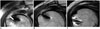
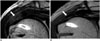
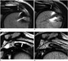
Two radiologists, who had 16 and 5 years experience in musculoskeletal imaging, independently reviewed the 100 postoperative MRIs from 50 patients (two follow-up MRIs from each patient) by consensus. The classification of SI patterns of the repaired tendons on T2-weighted scans is shown in Figure 1. Type I tendons showed heterogeneously high SI with fluid-like bright high SI foci (Fig. 1A), type II tendons exhibited heterogeneously high SI without fluid-like bright high SI foci (Fig. 1B), and type III tendons showed heterogeneously or homogeneously low SI (Fig. 1C). Tendon thickness was measured at the thickest portion of the tendon near the repair site below the acromion on oblique coronal scans (Fig. 5A, C). Additionally, the thickness of the rotator cuff interval was assessed by measuring the longest distance of the abnormal, ill-defined low SI between the coracohumeral ligament and the humeral head on oblique sagittal scans (Fig. 5C, D). Fluid in the intra-articular and periarticular (including the subacromio-subdeltoid space) spaces was assessed. Interval changes in these four findings were compared using the first and second postoperative MRI scans.
Finally, the distribution of the SI patterns of the 100 MRI scans (two postoperative MRI scans for each of the 50 patients), was evaluated individually without regard to serial scans, based on the follow-up period.
Statistical Analysis
The statistical analysis was performed using SPSS ver. 18.0 software for Windows (SPSS Inc., Chicago, IL, USA). The McNemar test was used to determine changes in the SI patterns of the repaired tendons between the first and second postoperative MRIs. The paired t test was used to determine differences in the thickness of repaired tendons and the rotator cuff interval. Data are expressed as mean ± standard deviation. A p value < 0.05 was considered significant.
RESULTS
Signal intensity patterns of the repaired tendons and their interval changes are summarized in Table 1. Heterogeneously high SI patterns, which corresponded to type I or type II, were observed in 45 tendons (90%) on the first MRI (type I, n = 20; type II, n = 25).
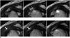
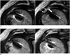
Of these 45 tendons, 30 (30/45, 66.7%) showed reduced SI on the second MRI. This was evident in 17 of 20 (85%) type I tendons (Figs. 2, 3) and in 13 of 25 (52%) type II tendons (Fig. 4). This pattern of decreased SI on the second MRI was significant (p < 0.050, McNemar test). Notably, of the 20 type I cases exhibiting fluid-like bright high SI, 11 changed to a type II pattern (Figs. 2, 5), and six cases changed to a type III pattern (Fig. 3).
Five tendons (10%) that had shown a type III pattern on the first MRI were consistently classified as type III on the second MRI. However, tendons with an unchanged SI pattern (three type I, 12 type II, and five type III) showed a similar tendency toward a decrease in SI, but were within the same SI pattern.
The distribution of SI patterns based on the follow-up period is shown in Figure 6, with a decreased incidence of the type I pattern in cases with longer follow-up periods, particularly those > 6 months.
Additionally, all type I and type II tendons classified on the first MRI contained one or two linear or curvilinear structures with intermediate to low SI, which appeared to be suture material, as well as small and ill-defined material within high SI areas at the repair site (Figs. 2A, 3A). These linear or curvilinear structures became partially or completely obscured by the decreased SI of the repaired tendons on the second MRI.
Forty-seven tendons (94%) showed decreased thickness over time (Fig. 5), with a mean thickness of 5.69 ± 1.34 mm on the first MRI and 4.69 ± 1.23 mm on the second MRI (p < 0.050) (Table 2).
The rotator cuff interval also became thinner on the second MRI (Fig. 5) in 35 patients (70%), with a mean thickness of 4.92 ± 1.47 mm on the first MRI and 3.64 ± 1.36 mm on the second MRI (p < 0.050) (Table 2).
The volume of periarticular or intra-articular fluid decreased in 38 cases (76%) (Fig. 5), showed no change in nine cases (18%), and increased in three cases (6%).
DISCUSSION
The postoperative status of a repaired tendon is evaluated using clinical assessments as well as imaging studies, such as plain radiographs, US, or MRI. Persistent shoulder pain after rotator cuff tendon repair may occur due to a variety of causes, such as tendinitis, persistent impingement, muscle atrophy, deltoid detachment, or tendon retear (6, 7, 8). Due to the difficulty formulating a differential diagnosis for clinically similar symptomatic manifestations, radiological evaluations play an important role in these cases. MRI is useful to evaluate the integrity of a repaired tendon as well as distinguish it from other potential causes of shoulder symptoms (2, 6, 9, 10, 11, 12, 13, 14).
Previous studies have shown that the appearance of a postoperative rotator cuff tendon can vary, including increased SI and thinning or contour irregularities in the tendon. These findings are regarded as tendinosis or degeneration in untreated tendons, but repaired tendons may appear normal postoperatively (2, 6, 9).
Postoperative MRI findings after rotator cuff repair primarily focused on the integrity of the repaired tendon, as previous studies have reported retear incidences of 5-94% (15, 16, 17, 18, 19, 20). This wide range may be related to MRI interpretation as well as patient factors, such as postoperative clinical outcome, preoperative tendon status (tear size, retraction degree, or patient age), or the surgeon's skill. Moreover, no study has found a correlation between tendon integrity on MRI and clinical outcome (4, 10, 21). Based on the above studies, orthopedic surgeons do not routinely perform second-look operations for repaired tendons with positive MRI findings but no symptoms. Notably, many surgeons avoid obtaining a postoperative MRI after rotator cuff repair due to the high cost and likelihood of a false-positive result, and this is one of the reasons why our study included only limited MRI sequences, which incurred a relatively low cost.
Accurate interpretation of a postoperative MRI requires an understanding of the normal postoperative MRI features of repaired tendons and the ability to distinguish them from retears. Spielmann et al. (22) analyzed MRI findings of patients with no postoperative symptoms and found that only 10% of patients showed normal tendon SI. Our study, which included patients clinically improving postoperatively, also showed that only five patients (10%) exhibited low SI on the first postoperative MRI. These results suggest that postoperative tendons must be evaluated differently from native tendons.
The diagnostic criteria for a retear in all previous postoperative MRI studies were either fluid SI foci replacing the tendon or a tendon defect on a T2-weighted image (2, 9). Such diagnostic criteria may make it difficult to distinguish a partial thickness retear from an edematous change (2). Some reports assert that such difficulty can be resolved with MR arthrography (23, 24). However, MR arthrography is unreliable due to possible leakage of contrast material that can occur in a well-repaired tendon and invasiveness (14). Some investigators have indicated that increased SI on T2-weighted images may cause a misdiagnosis as a retear due to postoperative hyperemic granulation tissue, edematous changes, or ongoing inflammation at the repair site (6, 9, 22). This suggestion has been supported by follow-up studies reporting interval changes in MRI SI of repaired tendons (4, 25, 26). Jost et al. (26) reported that eight of 20 patients diagnosed with a retear on the first postoperative MRI had no evidence of a retear on a long-term follow-up MRI 7.6 years later. Crim et al. (4) reported that tendon irregularities and thinning or increased SI was observed during the first postoperative year. Thus, such findings would be inappropriate to use as simple indicators of surgical repair failure. Although most MRIs in our study were performed within the first postoperative year (mean follow-up lengths for the first and second MRIs were 4.4 and 10.4 months, respectively), 66.7% of the repaired tendons with increased SI on the first MRI showed reduced SI on the second MRI. The type I pattern detected on the first MRI decreased in SI in 20 cases, including six cases that became nearly normal (type III pattern) on the second MRI.
Postoperative findings of other tendons have been evaluated. Serial MRIs after uncomplicated Achilles tendon repair show an inevitable tendon gap during the early follow-up period, although the incidence decreased over time and was absent 12 weeks after surgery (25). The authors suggested that this may have been related to the normal healing process. Similarly, our results suggest that increased SI with or without fluid-like bright high SI foci seen on an early follow-up MRI may not be a pathological finding but rather an early stage of the normal postoperative healing process in patients without remarkable symptoms. In addition, the linear or curvilinear structures and ill-defined debris-like material, which were observed within repaired high SI tendons, suggest that they were suture material within immature granulation tissue and the pseudomembrane along the repaired tendon.
Our results show that tendon thickness decreased significantly on the second follow-up MRI, indicating a decrease in edema over time. Similarly, thickness of the rotator cuff interval decreased significantly on the second follow-up MRI, suggesting decreased inflammation and edema.
Subacromio-subdeltoid bursal fluid is a common ancillary finding in cases of unrepaired tendon tears (2, 27, 28, 29). However, this is a relatively common finding that may develop after bursal resection or from bone marrow fluid leakage after acromioplasty or successful rotator cuff repair (9, 22, 30). In our study, the volume of periarticular or intra-articular fluid decreased in 38 cases and increased slightly in three cases on the second follow-up MRI. None of the three cases showed a retear, their SI patterns were type II or type III, and none showed any changes on the second follow-up MRI.
Several limitations to our study should be mentioned. First, given our retrospective study design, the follow-up period for each patient was inconsistent. However, our results indicate that frequency of the type III pattern tended to increase later in the follow-up, and that the incidence of the type I pattern was very low after postoperative month 6. Second, we only evaluated T2-weighted images of repaired tendons. However, T2-weighted images are sufficient to visualize continuity of a repaired tendon and adjacent fluid collection. Third, we did not obtain surgical confirmation for the postoperative MRI findings, as a second-look operation was rarely performed unless the patient was symptomatic. Thus, the MRI appearances including high SI of a repaired tendon could not be correlated with surgical or histological findings. Last, we did not compare MRI findings with those of patients who had a retear or remarkable clinical symptoms.
In conclusion, repaired supraspinatus tendons exhibited high SI on early postoperative MRIs performed 4.4 months after surgery in 90% of clinically improving patients. The high SI areas were characterized by linear or curvilinear structures and ill-defined intermediate SI debris. The increased SI and thickness of repaired tendons decreased on the later MRI. High SI or fluid-like bright high SI in repaired supraspinatus tendons on early postoperative MRIs in clinically improving patients suggests an ongoing healing process rather than a retear.


 XML Download