

 PDF
PDF ePub
ePub Citation
 Print
Print


INTRODUCTION
Coronary artery imaging in children is frequently challenging due to small size, high heart rates, and motion artifacts from cardiac pulsation, respiration, and the patients themselves, which results in technical or procedural difficulties (1). Imaging modalities for evaluating coronary arteries include catheter angiography, echocardiography, CT, and MRI. Awareness of the pros and cons of each imaging modality minimizes patient risks and maximizes diagnostic yields. Recent technical advancements in CT and MRI for evaluating the coronary arteries are extremely valuable (1, 2, 3).
Coronary artery imaging in children is important because the coronary blood flow to the myocardium and the cardiac conduction tissue maintains normal cardiac function. Various congenital and acquired coronary artery abnormalities in children, such as coronary artery anomalies, surgically important coronary artery anatomy specific to congenital heart disease, coronary artery abnormalities in Kawasaki disease, and cardiac allograft vasculopathy, can be identified with these imaging modalities. Review of imaging modalities in children for coronary artery, resultant coronary artery visibility, and clinical applicability will facilitate the practical clinical use of pediatric coronary artery imaging.
Imaging Modalities
High temporal resolution for high heart rates, high spatial resolution for small size, and low patient risks are required for coronary artery imaging in children (1, 2). The advantages and shortcomings of imaging modalities used for coronary artery imaging in children, including catheter angiography, echocardiography, CT, and MRI, were described in this section. Recent technical advancements in coronary CT angiography and coronary MR angiography were also reviewed.
Catheter Angiography
Catheter angiography has been used for evaluating coronary arteries in children for the past few decades. Aortography is generally used to visualize coronary arteries in small children younger than 1 year of age, while selective coronary angiography may be performed in older children (e.g., older than 5 years of age) (1). Complications associated with selective coronary angiography are not trivial, ranging from relatively frequent minor ones including temporary electrocardiography (ECG) changes (11.0%), transient bradycardia (2.5%), and vascular access-related complications (11.6%), to rare serious ones, such as ventricular fibrillation (0.6%) (4). Besides these complications, the use of ionizing radiation and iodinated contrast agent in catheter angiography is another concern particularly in children. Therefore, it should be reserved for interventional procedures or when noninvasive diagnostic imaging is inconclusive. Furthermore, because it is a 2-dimensional projection imaging, catheter angiography lacks the 3-dimensional spatial relationship between the coronary arteries and adjacent cardiovascular structures.
Echocardiography
Transthoracic 2-dimensional echocardiography along with color and pulsed-wave Doppler is widely used as the primary imaging modality of coronary arteries in children (5). However, echocardiography is often limited by operator dependency, poor acoustic window especially in older or postoperative children, and intrinsic diagnostic pitfalls (Fig. 1). For instance, an echo free space between the left atrial appendage and periaortic tissue may give a false impression of the left coronary artery normally arising from the left aortic sinus of Valsalva in children with anomalous origin of the left coronary artery from the pulmonary artery (ALCAPA) (5). As a result, confirmatory imaging, such as CT or MRI, should be performed if echocardiographic findings are equivocal or negative in patients with high clinical suspicion of coronary artery abnormalities.
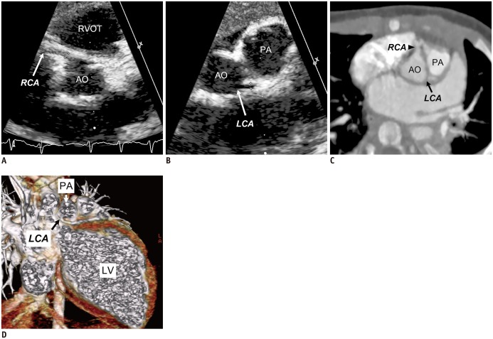 | Fig. 13-month-old boy with anomalous origin of left coronary artery (LCA) from pulmonary artery.Transthoracic echocardiographic images (A, B) show normal origins of right coronary artery (RCA, arrow in A) and LCA (arrow in B). C. Oblique axial cardiac CT image demonstrates normal origin of RCA (arrowhead) but no connection (arrow) of LCA to left aortic sinus. D. Cropped oblique coronal volume-rendered CT image clearly reveals anomalous origin (black arrow) of LCA from pulmonary trunk (PA) (white arrow). Dilated left ventricle (LV) is noted. AO = ascending aorta, RVOT = right ventricular outflow tract
|
CT
Coronary CT angiography is currently regarded as the diagnostic imaging method of choice for evaluating coronary arteries. ECG-synchronized scanning, i.e., either retrospective ECG gated spiral scanning or prospective ECG-triggered sequential scanning, should be performed for coronary CT angiography (2). To maximize the diagnostic yield of coronary CT angiography, selection of the best cardiac phase and the optimal scan delay with an optimized contrast injection protocol is crucial (2, 6, 7, 8). The recently introduced high-pitch dual-source spiral scanning with prospective ECG triggering enabling the whole scan in one heartbeat may also be used for evaluating coronary arteries (6, 7). It should be noted that images acquired with this scan mode show different cardiac phases along the z-axis, which is clearly different from ECG-synchronized scans showing the same cardiac phase for the entire scanned volume. Although the image quality of ECG-triggered high-pitch dual-source spiral scan is obviously inferior to those of ECG-synchronized scans, it serves as a troubleshooting scanning technique in irritable or unstable children (Fig. 2). As beta-blockers or sublingual nitroglycerin is frequently employed prior to coronary CT angiography to decrease the heart rate or to dilate the coronary arteries in adults, these preparations should be carefully applied to children because beta-blockers substantially prolong patient preparation time with no guaranteed success and the unpleasant taste of sublingual nitroglycerin may irritate pediatric patients (9). In addition to coronary artery morphology, CT can show first-pass myocardial hypoperfusion and delayed myocardial enhancement resulting from significant coronary artery stenosis or occlusion, as in cardiac MRI (10). However, optimal imaging protocol and clinical value of delayed myocardial enhancement CT imaging have yet to be established in children (Fig. 3). Minimal CT radiation dose while maintaining diagnostic image quality can be achieved by various radiation dose-reduction strategies, such as individual body size-adapted scan parameters, minimal longitudinal scan range, tube current modulation(ECG-controlled or not), low tube voltage, adaptive section collimation, and iterative reconstruction algorithm, which is of utmost importance in children (2, 6, 7, 11, 12, 13, 14, 15). It is noteworthy that pediatric cardiac CT is more frequently used than pediatric cardiac MRI in Asian countries for several reasons (16).
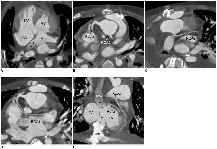 | Fig. 211-year-old boy who underwent double switch operation for congenitally-corrected TGA.
A. 4-chamber cardiac CT image shows morphologic left ventricle (LV), morphologic right ventricle (RV), morphologic right atrium (RA), morphologic left atrium (LA), and patent systemic venous baffle (asterisks). Oblique axial (B-D) and coronal (E) cardiac CT images demonstrate patent transferred coronary arteries, dual RCA (RCA1, RCA2), and patent systemic venous baffle (asterisks). IVC = inferior vena cava, RCA = right coronary artery, SVC = superior vena cava, TGA = transposition of great arteries
|
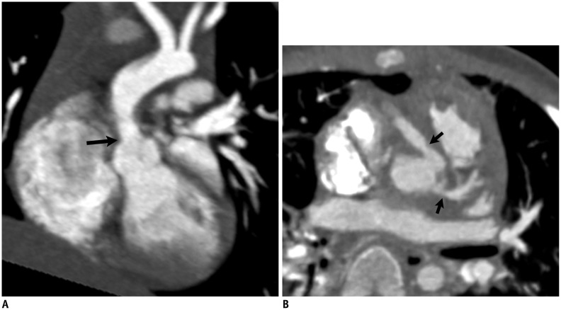
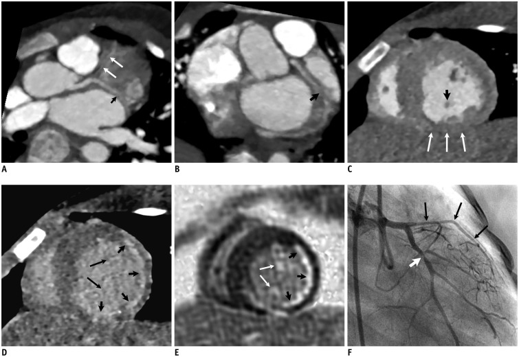 | Fig. 33-year-old boy with cardiac involvement of chronic graft-versus-host disease after hematopoietic stem cell transplantation for chronic granulomatous disease.Cardiac CT images (A, B) show long-segment severe narrowing (long arrows) of left anterior descending artery and focal narrowing (short arrow) of proximal left circumflex artery. C. Early short-axis cardiac CT image reveals thinning of inferior wall (long arrows) and posteromedial papillary muscle (short arrow) of left ventricle. D. 6-min-delayed short-axis cardiac CT image demonstrates subendocardial myocardial delayed enhancement (short arrows) in left coronary artery territory and left ventricular papillary muscles (long arrows) indicating myocardial infarction, which is in accordance with short-axis late gadolinium enhancement MR image (E). F. Catheter left coronary angiography confirms long-segment severe narrowing (long arrows) of left anterior descending artery and focal narrowing (short arrow) of proximal left circumflex artery as seen on cardiac CT (A, B).
|
MRI
Free-breathing, ECG-triggered, navigator-gated, T2-prepared, 3-dimensional coronary MR angiography using steady state free precession (SSFP) sequence, is the current standard in clinical practice. The imaging technique does not require the administration of contrast agent due to endogenous contrast between the coronary arteries and the surrounding tissue, accentuated by fat saturation and T2 preparation prepulses. However, examination using the SSFP sequence is often performed after the injection of a gadolinium-based extracellular contrast agent for other reasons, such as contrast-enhanced MR angiography and late gadolinium enhancement MRI because the signal-to-noise ratio is considerably increased on the contrast-enhanced study at either 1.5-T or 3.0-T (Fig. 4). Although it does not use ionizing radiation, coronary MR angiography is limited by long examination time and low spatial resolution that are problematic in imaging young children. Coronary CT angiography, only requires adjustment of the trigger delay to obtain the best cardiac phase because the duration of the acquisition window, i.e., the temporal resolution, is fixed. In contrast, the 2 parameters are independently determined according to the cardiac rest periods evaluated with 50-80-phase 4-chamber cine imaging for each patient in coronary MR angiography. Depending on the data acquisition method of coronary MR angiography, 2 approaches, i.e., target-volume vs. whole-heart, are available (Fig. 5) (3, 9). Multiple sub-volumes targeted on each coronary artery are obtained in the target-volume approach, while only 1 volume, usually in the axial plane, including the whole heart is acquired for the whole-heart approach. The image quality is the same between the 2 approaches (3). As compared with the whole-heart approach, the main drawbacks of the target-volume approach include limited volumetric coverage, low visibility of the coronary artery branches, and operator dependency in localizing the scan plane. Multi-planar and 3-dimensional visualization of all the 3 major coronary arteries is an advantage of the whole-heart approach with isotropic voxels. Shorter imaging time in coronary MR angiography has a higher success rate. This can be achieved by using a 32-channel cardiac coil and a high parallel imaging factor. Recent studies (3, 17) show that inversion-recovery SSFP sequence with an intravascular blood pool contrast agent, such as gadofosveset trisodium, substantially improves image quality and diagnostic performance of coronary MR angiography compared with T2-prepared SSFP sequence. However, gadofosveset trisodium is not widely available in Asia.
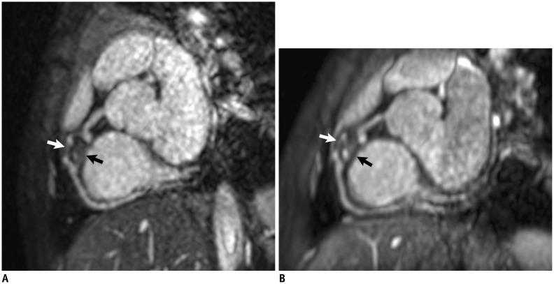
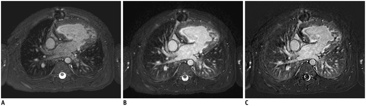 | Fig. 43-year-old boy with functional single ventricle who underwent extracardiac conduit Fontan operation.Pre- (A) and post-contrast (B) navigator-gated coronary MR angiographic images obtained at 1.5-T MR scanner demonstrate that cardiovascular structures are substantially enhanced after intravenous administration of extracellular gadolinium-based contrast agent. C. Subtraction image clearly show effect of contrast-enhancement in coronary MR angiography. Normal enhancement of dependent portions of both lungs is also noted (B, C).
|
Abnormally thickened coronary artery walls can be imaged by using an ECG-triggered, navigator-gated double inversion recovery black-blood segmented turbo spin-echo sequence (18, 19). However, long acquisition time limits its practical use. Moreover, slow flow artifacts indistinguishable from mural thrombus or eccentric wall thickening should be recognized as a diagnostic pitfall of the coronary vessel wall MRI (Fig. 6). In contrast to CT, cardiac MRI can provide comprehensive assessment of cardiac function, myocardial perfusion, and myocardial viability as well as coronary artery morphology (9, 20, 21).
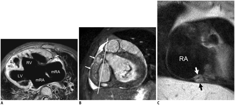 | Fig. 6Coronary vessel wall MRI.
A. Axial electrocardiography (ECG)-triggered, navigator-gated, double inversion recovery, fat-saturated black-blood segmented turbo spin-echo MRI in 3-year-old girl with functional single ventricle and right isomerism who underwent bidirectional cavopulmonary shunt shows wall (arrows) of normal right coronary artery. In addition, dextrocardia, large ventricular septal defect, and remnant of interatrial septum are noted. C. Oblique coronal ECG-triggered, navigator-gated, double inversion recovery, fat-saturated black-blood segmented turbo spin-echo MRI obtained along line in B in 3-year-old boy with Kawasaki disease and thrombosed fusiform aneurysm (long arrows in B) of right coronary artery reveals severe concentric wall thickening (short arrows in C) of distal right coronary artery. LV = left ventricle, mRA = morphologic right atrium, RA = right atrium, RV = right ventricle
|
Coronary Artery Visibility in Children
Coronary artery visibility on CT and MRI tends to be compromised in young children due to the small size of the coronary arteries and very high heart rates. Nevertheless, coronary artery visibility has markedly improved with recent technical improvement in CT and MRI (Table 1) (9, 20, 22, 23, 24, 25, 26, 27). ECG-synchronized CT scanning for coronary arteries should be used in children because the scan technique shows significantly higher coronary artery visibility than non-ECG-synchronized CT scanning (Table 1). In children, coronary artery visibility for all segments on ECG-synchronized CT largely ranges from 79% to 89%, which is higher than that (60-79%) on MRI (Table 1). Despite intensive imaging procedures, such as general anesthesia with repeated ventilator stoppings, the success rate of coronary MR angiography is merely 17% in infants younger than 4 months of age (27). However, the success rate of coronary CT angiography is substantially higher than coronary MR angiography in newborns and infants (23, 24, 25). In addition, coronary CT angiography is generally better than coronary MR angiography in evaluating coronary luminal stenosis in children, primarily due to higher spatial resolution of CT.
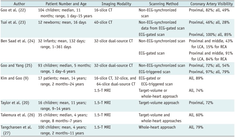
Table 1
Coronary Artery Visibility on CT and MRI in Children
| Author | Patient Number and Age | Imaging Modality | Scanning Method | Coronary Artery Visibility |
|---|---|---|---|---|
| Goo et al. (22) | 104 children; median, 11 months; range, 1 day-15 years | 16-slice CT | Non-ECG-synchronized scan | Proximal, 82%; all, 49% |
| Tsai et al. (23) | 12 newborns; mean, 16 days | 40-slice CT | Non-ECG-synchronized data from ECG-gated scan | Proximal, 46%; all, 28% |
| ECG-gated scan | Proximal, 100%; all, 85% | |||
| Ben Saad et al. (24) | 32 infants; mean, 132 days; range, 1-361 days | 32-slice dual-source CT | Non-ECG-synchronized scan | Proximal and middle, 43% for LCA, 15% for RCA |
| ECG-gated scan | Proximal and middle, 91% for LCA, 84% for RCA | |||
| Goo and Yang (25) | 93 children; median, 5 months; range, 1 day-6 years | 32-slice dual-source CT | Non-ECG-synchronized scan | Proximal, 72%; all, 54% |
| ECG-triggered scan | Proximal, 97%; all, 79% | |||
| Kim and Goo (9) | 17 patients; mean, 14 years; range, 2 months-24 years | 16-slice CT, 32-slice, and 64-slice dual-source CT | ECG-gated or ECG-triggered scan | All, 89% |
| 1.5-T MRI | Target-volume or whole-heart approach | All, 74% | ||
| Taylor et al. (20) | 16 children; mean, 11 years; range, 9-14 years | 1.5-T MRI | Target-volume approach | Proximal, 72% |
| Takemura et al. (26) | 35 children; median, 4 years; range, 8 months-7 years | 1.5-T MRI | Target-volume and whole-heart approaches | All, 60% |
| Tangcharoen et al. (27) | 100 children; mean, 4 years; range, 2 months-11 years | 1.5-T MRI | Whole-heart approach | All, 79% |
![]()
The selection of the best cardiac phase is required to increase coronary artery visibility equally on both CT and MRI. The mid-diastolic phase usually provides the best image quality of the coronary arteries at slow and regular heart rates, though such heart rhythms are uncommon in children. At higher heart rates (e.g., > 70-75 bpm), the end-systolic phase is apt to offer higher coronary artery visibility than the mid-diastolic phase because the mid-diastolic cardiac rest period gets invariably shorter with increasing heart rates. The end-systolic phase is more resistant to arrhythmia or heart rate variability than other cardiac phases. Although not fully recognized yet, the end-diastolic phase may be used as the third alternative for the best cardiac phase.
Clinical Applications
With recent improvements in coronary artery visibility on CT and MRI in children as described above, the diagnostic accuracy and the resultant clinical utility of coronary CT angiography and coronary MR angiography are noticeably increasing in children. Hence, we need to recognize the extent to which we can enhance pediatric coronary artery imaging with the recent technical improvements in clinical practice. We accordingly described various congenital and acquired coronary artery abnormalities in children in this section.
Coronary Artery Anomalies
Coronary artery anomaly is defined as an anatomical variation observed in < 1% of the general population. Two classification systems for coronary artery anomalies are commonly used (28, 29). One is according to the location of the anomalies, including origin, course, and termination, while the other is according to hemodynamic significance of the anomalies. Hemodynamically significant coronary artery anomalies may compromise myocardial perfusion that leads to adverse cardiac events, such as myocardial ischemia, myocardial infarction, or sudden cardiac death, in children. They include atresia of the left coronary artery, ALCAPA, anomalous origin of a coronary artery from the opposite sinus of Valsalva with interarterial/intramural course, and coronary artery fistula. Atresia involving the left main coronary artery is most often described (29). However, the right coronary artery (Fig. 7) or the left circumflex artery may be rarely affected (29). Collateral vessels are usually developed from patent coronary artery to the atretic coronary artery (Fig. 7). However, the collateral flow is generally insufficient to meet the oxygen requirement of the left ventricular myocardium occurring mostly in the first year of life in atresia of the left main coronary artery. ALCAPA usually presents with congestive heart failure in infants and should be distinguished from dilated cardiomyopathy at this age (30). Because echocardiographic findings may be often inconclusive (5), CT or MRI can be used for identifying ALCAPA and the extent of myocardial infarction (Fig. 1). A recent study (31) showed that intramural segments in anomalous coronary arteries with interarterial course can be identified on coronary CT angiography by demonstrating acute angle of origin, slit-like orifice, and elliptical cross-sectional shape with vessel height/width ratio greater than 1.3. Coronary artery fistula is defined as a direct precapillary connection between a coronary artery and a low-pressure system, such as the right cardiac chamber, pulmonary artery, or coronary sinus, resulting in a usually small left-to-right shunt (32, 33, 34, 35). Coronary artery fistula is generally asymptomatic in adults, while it is often symptomatic in pediatric patients. The majority of coronary artery fistulas have single communications and most symptomatic fistulas arise from the right coronary artery. Coronary artery fistula usually occurs in isolation but may occur in complex congenital heart disease, such as pulmonary atresia and intact ventricular septum in which myocardial ischemia may be developed due to the so-called right ventricle-dependent coronary circulation (35). Coronary CT angiography provides detailed angioarchitecture of coronary artery fistula and its spatial relationship with the nearby major cardiovascular structures, which is necessary for optimal treatment planning (32, 33, 34, 35). In addition to hemodynamically significant anomalies, hemodynamically benign coronary artery anomaly, such as high take-off, may be clinically significant at cardiac surgery (28, 36) (Fig. 7).
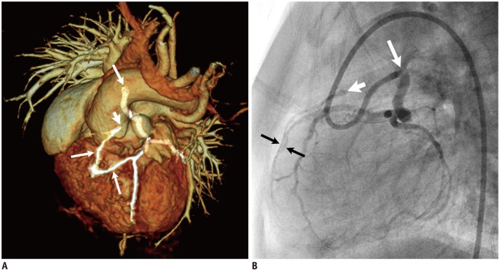 | Fig. 79-month-old girl with tetralogy of Fallot who underwent left Blalock-Taussig shunt.Volume-rendered cardiac CT image (A) and lateral view of catheter left coronary angiography (B) demonstrate high take-off (long arrow) left coronary artery, atresia (short arrow) of right coronary artery, and collateral artery between left and right coronary arteries (long arrows).
|
Congenital Heart Disease
Cardiac CT is increasingly used for preoperative morphologic evaluation of congenital heart disease (2, 6, 7, 16, 37, 38, 39). Coronary arteries are commonly included in the evaluation due to recently improved coronary artery visibility on cardiac CT (23, 24, 25). Prevalence of coronary artery anomalies in patients with congenital heart disease is higher than that in the general population. In addition, certain coronary artery anatomy should be vigilantly depicted on CT or MRI in certain congenital heart disease, such as transposition of the great arteries or tetralogy of Fallot, to avoid potential damage to these aberrant coronary arteries during or after surgery (27, 28, 40, 41). Moreover, the left main coronary artery may rarely be extrinsically compressed by a dilated pulmonary artery in various congenital heart diseases or pulmonary arterial hypertension, as demonstrated on catheter coronary angiography or coronary CT angiography (42, 43, 44). The affected left coronary artery is possibly vulnerable to the extrinsic compression because it tends to originate from the right side of the left aortic sinus. Postoperative CT or MRI can also be used to identify significant coronary artery stenosis and to elucidate the underlying mechanism, i.e., stretching, kinking, or compression, of coronary luminal narrowing in children with congenital heart disease (20, 28, 45). Coronary CT angiography is useful to evaluate native coronary artery patency, as well as bypass graft patency in children with congenital or acquired heart disease after coronary revascularization surgery (Fig. 8) (46).
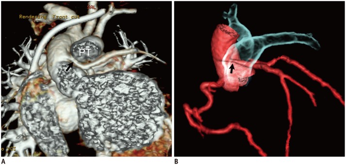 | Fig. 85-month-old girl who underwent reimplantation of anomalous left coronary artery from pulmonary artery.
A. Cropped coronal volume-rendered cardiac CT image shows extrinsic compression (arrow) of reimplanted left main coronary artery for pulmonary trunk (PT) that is better depicted on segmented volume-rendered image of coronary artery and pulmonary artery (B).
|
Kawasaki Disease
Kawasaki disease is the most common cause of acquired coronary artery disease in children necessitating lifelong imaging surveillance for coronary artery obstruction and altered myocardial perfusion (9, 18, 21, 26, 47, 48, 49, 50, 51, 52, 53). Echocardiography is the primary imaging modality to identify a subset (approximately 15-25%) of patients with coronary artery dilatation or aneurysm during the acute phase of the disease up to the first 6 weeks after presentation. At this period, CT or MRI may be used for complete initial mapping of the coronary artery abnormalities required for accurate risk stratification because echocardiography is often limited in evaluating middle and distal segments of the coronary arteries, particularly in growing children. These coronary artery aneurysms are then gradually subject to regression, stenosis, occlusion, thrombosis, and myointimal thickening, frequently without overt clinical manifestations (Figs. 5, 6). For serial imaging follow-up, assessment of myocardial perfusion is generally recommended in addition to morphologic assessment of the coronary arteries. Myocardial perfusion scintigraphy may be omitted and 1-stop shop evaluation is feasible when stress myocardial perfusion imaging with CT or MRI is added to the cardiac imaging protocol for patients with Kawasaki disease. For evaluating coronary luminal stenosis, coronary CT angiography appears slightly better than coronary MR angiography (9, 49). The benefit-risk ratio of coronary CT angiography becomes more favorable with various radiation dose-reducing techniques. Serial follow-up coronary CT angiography at a 3-year interval in patients with Kawasaki disease is regarded as acceptable by our institution, but there are no established guidelines yet. Optimal diagnostic algorithm, for evaluating coronary artery abnormalities in Kawasaki disease based on an expert consensus opinion, remains to be updated with the recent technical advances in cardiac CT and MRI. As a result, catheter coronary angiography can be reserved when noninvasive imaging findings are inconclusive or equivocal, or when interventions are planned.
Williams Syndrome
Supravalvular aortic stenosis and peripheral pulmonary artery stenosis are a hallmark of cardiovascular manifestations of Williams syndrome (54, 55). Supravalvular aortic stenosis is associated with an increased risk of sudden cardiac death in these patients. The so-called "coronary hooding" phenomenon occurred by the aortic valve leaflets fused to the sinotubular junction may result in myocardial ischemia. Furthermore, the coronary arteries located proximal to the supravalvular aortic stenosis are subject to high systolic pressures and result in coronary artery stenosis. Cardiac CT is useful to evaluate cardiovascular manifestations, including supravalvular aortic stenosis and coronary artery abnormalities, of Williams syndrome (Fig. 9) (54, 55).
Cardiac Allograft Vasculopathy
Cardiac allograft vasculopathy is one of the late complications of heart transplantation, typically seen as a diffuse, concentric, noncalcified wall thickening of the coronary arteries (19, 47, 56, 57). The affected patients are usually clinically asymptomatic because the transplanted heart often remains denervated. Consequently, imaging surveillance using accurate and noninvasive techniques is needed. Coronary CT angiography is regarded as the imaging method of choice for the diagnosis of cardiac allograft vasculopathy because it has a high sensitivity, specificity, and negative predictive value comparable to invasive catheter coronary angiography (56, 57). A 2-year follow-up interval appears to be safe for detecting a significant coronary artery lesion when the previous coronary CT angiography is normal (56). On the other hand, late gadolinium enhancement MRI of the coronary vessel wall may be used to detect and grade cardiac allograft vasculopathy (19).
Cardiac involvement of graft-versus-host disease is very rare and one of the major causes of morbidity and mortality after hematopoietic stem cell transplantation (58). Similar to cardiac allograft vasculopathy, multiple concentric narrowings of the coronary arteries may be developed in patients with cardiac graft-versus-host disease (Fig. 3). Bradycardia or cardiomyolysis may also occur (58).
Go to : 
CONCLUSION
After recognizing the strengths and weaknesses of imaging modalities for coronary artery imaging, we can optimize imaging protocols and algorithms for evaluating and monitoring congenital and acquired coronary artery abnormalities in children. CT and MRI play a key role in overcoming the diagnostic challenges in coronary artery imaging in children by avoiding the diagnostic pitfalls of echocardiography and reducing risks related to catheter coronary angiography. Coronary CT angiography is particularly useful in newborns and young infants.
Go to :
 XML Download