

 PDF
PDF ePub
ePub Citation
 Print
Print


INTRODUCTION
Bone age assessment allows the evaluation of developmental status, sexual maturity and prediction of the ultimate height of children. Bone age assessment plays an important role in diagnosis and monitoring of treatment of growth disorders and endocrinological abnormalities in children by detecting significant discrepancy between bone age and chronological age (1).
For evaluation of bone age, two major methods are generally used globally in clinical practice based on radiograph of left hand-wrist: the Greulich-Pyle (GP) method (2) and the Tanner-Whitehouse 3 (TW3) method (3). Both methods have been used to assess bone age in Korean children (4, 5), although whether these standards can be applied to the current assessment of bone age in children of different ethnicities is questionable. In 1999, Korean standard bone age chart (KS) was published, which was derived from radiographs of 3407 Korean children (6, 7). Since then, the KS has been widely used in Korea.
There have been some comparative studies between the GP method and TW3 method around the world including Korea (8,9,10,11,12). But, no study about KS compared to the GP or TW3 method has been done. Therefore, the aim of this study was to compare the reliability of these three methods (GP, TW3, and KS) in the evaluation of bone age of Korean children.
MATERIALS AND METHODS
We selected left hand-wrist radiographs of 278 prepubertal Korean children (aged between 7 and 12 years) between January 1, 2002 and December 31, 2012. These radiographs were obtained for the evaluation of the traumatic injury in the emergency department. Radiographs of 66 children were excluded from the study due to inadequate radiographic quality, poor position, bony abnormalities including fracture and those were derived from children who were diagnosed as growth disorders or endocrinological disorders. A total of 212 radiographs were analyzed, which included 135 boys and 77 girls.
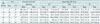
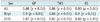
Bone age was estimated independently by two observers including one attending radiologist and one resident. Bone age was estimated using the GP method, TW3 method, and KS. The TW3 method was calculated on the average of the Radius-Ulna-Short bone (RUS) score and carpal score. When there were some mismatches between the patient's radiography and image of KS chart or GP atlas, we decided the mean value of two most similar images of KS chart or GP atlas as the patient's bone age. Patient-identifying information, except sex, was masked during interpretation including subject's chronological age. Bone age and chronological age were calculated in months (Table 1).
The correlation between chronological age and bone age measured by each method was analyzed using Pearson correlation coefficient and scatterplot. The differences between the chronological age and the measured bone ages by each method were calculated for each child, and one-way analysis of variance (ANOVA) was used to test of statistical significance of difference between three methods using those calculated values. Interobserver reliability coefficient was also calculated using intra-class correlation coefficient by each method. All analyses were performed using SPSS software for Windows version 17.0 (SPSS Inc., Chicago, IL, USA). P values < 0.01 were considered to be statistically significant.
RESULTS
In the whole group and the group of boys, the average of bone age estimated by KS was the closest to the average of chronological age. But, in girls, the average of bone age estimated by TW3 method was the closest to the average of chronological age (Table 2). Bone age measured by GP method tended to younger than chronological age in the whole group and the group of boys. On the other hand, bone age measured by TW3 method tended to older than chronological age in the whole group and in each gender.
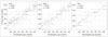
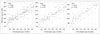
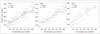
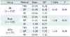
The correlations between chronological age and bone age estimated by each method are shown in Figures 1, 2, and 3. There were significant correlations between chronological age and bone age estimated by the three methods in the whole group and each gender (R2 ranged from 0.87 to 0.9, p < 0.01). Table 3 summarizes correlation coefficients between chronological age and bone age measured with each method. In the whole group and the group of girls, correlation coefficient between chronological age and bone age estimated by KS (R2 = 0.89 in the whole group and the group of girls) was slightly higher than others (GP and TW3, R2 = 0.88 in the whole group and 0.87 in the group of girls). In the group of boys, the correlation coefficient between chronological age and bone age estimated by KS and GP (R2 = 0.9) were slightly higher than TW3 (R2 = 0.88). But, according to the result of one-way ANOVA (Table 4), the difference between three methods was not statistically significant in the whole group (p = 0.9) and in each gender (p = 0.34 in boys and 0.29 in girls).
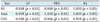
Average bone age differences among individual observers are given in Table 5. The average difference in bone age assessments among observers were 0.45 ± 1.79 months by the GP method, 0.45 ± 1.81 months by the TW3 method and 0.21 ± 1.19 months by the KS method. The interobserver reliability coefficients were more than 0.95 in all three methods (ranging from 0.996 to 1.000) (Table 6).
DISCUSSION
Bone age reflects current developmental status and sexual maturity of children and allows prediction of the ultimate height. Thus, assessment of bone age is important in the diagnosis and monitoring of the treatment of growth disorders and endocrinologic disorders.
There are various methods for bone age assessment, but two major methods are commonly used globally for bone age assessment in children based on radiography of left hand-wrist: the GP method (2) and the TW3 method (3). The GP method was derived from white children of upper socioeconomic level in United States of America during the 1930s and the second edition was published in 1959. Bone age determination is performed by comparing the left hand-wrist radiograph of a patient with the GP atlas, so it is relatively simple and convenient. The TW method was derived from British children in the 1950s and it was revised as TW3 method in 2001. The TW3 method uses a detailed shape analysis of several bones, and bone age assessment by TW3 method is performed by scoring each ossification centers and plotting the final sum. Thus, some researchers claim that the TW3 method provides high reproducibility and less intra- and inter-observer difference (10,11,12), but it is time-consuming and difficult to apply.
In Korea, the GP and TW3 methods are generally used for bone age estimation. However, some radiologists and clinicians have certain reservations regarding the applicability of these two methods in the evaluation of skeletal maturity of current Korean children, because subjects of these two methods have different ethnicities from Korean children, and these are long-established. A study by Ontell et al. (4) suggested GP method showed significant discrepancy in black and Hispanic girls, and in Asian and Hispanic boys in late childhood and adolescence. Also Zhang et al. (5) claimed that there were ethnic and racial differences in growth patterns at certain ages, and bone age measured with GP method was significantly overestimated in Asian and Hispanic children. Asian children tend to attain ultimate height earlier than Western children (13, 14).
Yeon and Kim (6, 7) established KS derived from radiographs of the left hand of 3407 healthy Korean children in 1999. As with the GP method, bone age estimation by KS is performed by comparing the left hand-wrist radiograph of child with KS chart, so it is easy to apply and convenient to use in the out-patient setting.
Despite the passage of more than 10 years since the KS was established, there has been no comparative study between GP, TW3, and KS. However there were several studies about comparison between GP method and TW3 method globally (8,9,10,11,12). Kim et al. (8) investigated the difference of bone age comparing GP and TW3 method, and there was a significant correlation between the GP and TW3 methods without significant difference of intraobserver variation in the bone age assessment of Korean children. So the authors claimed both GP and TW3 methods are useful for estimation of bone age in Korean children.
In this study, we compared the reliability of these three methods (GP, TW3, and KS) in the evaluation of bone age of healthy prepubertal Korean children. The correlations between those three methods and chronological age were strong. Therefore, the GP, TW3, and KS methods are well-suited for the evaluation of the bone age of healthy Korean children. KS showed higher correlation with chronological age than GP or TW3, but the difference between them was not significant.
There are several limitations to the current study. First, the number of subjects was small and age groups including adolescents and preschool age children were not included in the study. Furthermore, while intraobserver variation is important in the study about bone age estimation, this study did not include intraobserver variability.
In conclusion, the KS, GP, and TW3 methods are useful for evaluation of bone age in Korean children. For the KS to be used as the radiological index in the maturity status of Korean children, a multi-institutional study with a large population may be needed. As there are worldwide efforts to establish reliable computer-assisted bone age estimation, including computer-assisted skeletal age scores based on the TW3 RUS method, we should consider reinforcement and modification of the KS to make it an index of computer-assisted bone age estimation in Korean children.


 XML Download