

 PDF
PDF ePub
ePub Citation
 Print
Print


INTRODUCTION
The majority of sudden cardiac deaths are due to coronary plaque rupture. Myocardial infarction due to coronary atherosclerotic rupture is one of the main causes of mortality in young adults (1). Despite the recent advancements in various imaging modalities, identification of high-risk coronary plaque is still difficult. Anatomical imaging to assess luminal patency, such as computed tomography (CT) coronary angiography, have failed to detect high-risk plaque, because the vulnerable vessel normally has non-flowing limiting plaque, which is a high-risk plaque called "vulnerable plaque."
To overcome the limitations of anatomical imaging, several molecular imaging probes are currently being investigated to target biomarkers in vulnerable plaque. Molecular markers for atheromatous plaque can provide clinically crucial information regarding the vulnerability and the degree of progression. In addition, molecular imaging has a potential to permit the development of a novel therapeutic agents and non-invasive imaging tool to monitor the therapy response (23).
In this study, we reviewed the pathophysiology of calcification in atherosclerotic plaque and F-18 fluoride positron emission tomography (PET) as an imaging probe for vulnerable plaque.
Pathophysiologic Basis of Atherosclerotic Plaque
The pathogenesis of atheromatous plaque formation involves cholesterol deposition, macrophage accumulation, inflammation, and smooth muscle proliferation. The characteristics of vulnerable plaque include a large necrotic core, thin fibrous cap, inflammation, macrophage aggregation, hypoxia, and microcalcification (456). Inflammation is the key pathophysiology in the formation and progression of atherosclerotic plaque. Pathology confirmed macrophage aggregation within the plaque lipid core (4).
Early plaque calcification was observed in the thin fibrous cap overlying the necrotic core of atherosclerotic plaque. Studies have shown that microcalcifications in the thin fibrous cap increased the risk of plaque rupture, and subsequent stress-related microfractures result in acute thrombosis (78). Calcification is part of the healing process after inflammation. Similar to the calcification observed in tuberculosis patients, calcification occurs in patients with atheroma during the healing process after inflammation in the necrotic core (9).
Calcification in patients with atheroma is a bi-phasic reaction. The early phase is not visible on conventional imaging, but is associated with plaque instability. The latter phase of macroscopic calcification can be observed on a radiograph or CT (10). Normally, the diameter of microcalcification is small (< 5 mm), occurs during the early stage of calcification, has inflammation, covers a large surface area, has high exposed hydroxyapatite, and possesses a high risk for rupture (9).
The mechanism of vessel calcification is not well elucidated, but it is thought that vessel calcification in atheromatous plaque progression is an active process (11). Active vessel calcification is divided into 3 stages: initiation, propagation, and end-stage calcification (12). After prolonged inflammation from the initiation and propagation phases, microcalcification is formed in the plaque. Regional distribution of inflammation and microcalcification appears to overlap or is in close proximity to the initial in vivo imaging (13). Microcalcification may induce a pro-inflammatory response leading to plaque rupture by microcalcification fracture (14). The end-stage calcification phase manifests as advanced tissue calcification, which can be easily detected on CT and may be an irreversible process.
Current Imaging Method for Plaque Evaluation: FDG
Several biomolecules have been suggested as possible targets for atherosclerotic plaque. F-18 fluorodeoxyglucose (FDG) is a well-known marker for glucose metabolism. Because of high macrophage accumulation in the atheromatous plaque, the plaque has high FDG uptake, especially if is vulnerable (1516).
Feasibility of F-18 Fluoride PET in Atherosclerotic Plaque
Fluoride has a strong affinity for bone structures, and its radioactive isotope, F-18 fluoride, was proposed as a clinical imaging probe for skeleton imaging (17). However, after the wide-spread of single photon emission computed tomography camera, F-18 fluoride PET was replaced by Tc-99m labeled diphosphonate agents. Compared with bone scintigraphy using Tc-99m-labeled diphosphonate agents, F-18 fluoride PET has a unique advantage of high resolution. Due to high image quality and higher sensitivity than Tc-99m labeled diphosphonate agents, F-18 fluoride PET can be used for the evaluation of primary and metastatic bone tumors (18). Recently, the introduction of vascular PET imaging is feasible in many PET centers. Many cyclotron centers are producing F-18 fluoride for bone imaging, and F-18 fluoride PET already has been easily available for vascular imaging.
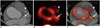
Recently, F-18 fluoride PET was suggested as a promising imaging probe for atheromatous plaque imaging. F-18 fluoride ions accumulated in bone-forming fluoroapatite crystal by exchange of hydroxyl groups on hydroxyapatite surface (19). Similar to F-18 fluoride deposits in various active calcification sites, these ions may accumulate in vulnerable plaque (Fig. 1). In advanced calcified plaque, which is stable, and in the advanced stage, no deposition of F-18 fluoride may be observed.
F-18 fluoride uptake in the aortic arterial wall was assessed in oncology patients who were under bone metastasis work-up (20). Out of all the patients assessed, 76% showed vascular wall F-18 fluoride uptake, with high uptakes in the femoral artery, abdominal aorta, and thoracic aorta. Analysis of the lesions showed that only 12% of arterial calcification sites had increased F-18 fluoride uptake. Most of the vascular calcification detected on CT should be regarded as stable, chronic stage of atherosclerosis, if there was no F-18 fluoride uptake, which is a marker for active microcalcification.
Derlin et al. (21) compared F-18 fluoride, FDG and CT calcification at major vessels in 45 oncologic patients, simultaneously. Common carotid artery, ascending aorta, aortic arch, descending aorta, and abdominal aorta were evaluated. The results revealed FDG uptake rates of 14.5%, while the F-18 fluoride level was 77.1% at CT calcification sites. Coincident uptake of both FDG and F-18 fluoride was observed in only 6.5%, thereby suggesting a difference in the biologic target of these two probes. It was suggested that FDG and fluoride may allow evaluation of distinct pathophysiologic processes in atherosclerotic lesions.
The carotid arterial F-18 fluoride uptake was assessed, and the significant correlation between F-18 fluoride uptake and CT calcification was determined (22). They evaluated 260 oncologic patients who underwent F-18 fluoride PET for bone metastasis evaluation. 94 patients (34.9%) of the patients had vascular F-18 fluoride uptake, and its uptake was colocalized with calcification in all atherosclerotic lesions. High fluoride uptake was associated with age, male, hypertension, and hypercholesterolemia. They reported a highly significant correlation between the F-18 fluoride uptake and number of cardiovascular risk factors. It was suggested that F-18 fluoride uptake may quantify continuing mineral deposition in carotid plaque, which suggest a potential role of F-18 fluoride for imaging and characterization of carotid atherosclerotic plaque.
The feasibility of F-18 fluoride PET to assess coronary artery was assessed in volunteers and patients with aortic stenosis by Dweck et al. (23). Focal F-18 fluoride uptake was observed in the coronary artery and could be determined using a hybrid CT image. There were discordant sites between coronary F-18 fluoride uptake and CT calcification. Coronary calcification without any F-18 fluoride uptake was frequently observed. Subjects (41%) with a high coronary calcium score (> 1000) did not show significant F-18 fluoride uptake. This finding suggested that F-18 fluoride PET provides different information relating to metabolically active calcific plaque and developing microcalcification. This study compared the F-18 fluoride uptake with FDG in the coronary artery. Coronary FDG PET images were not adequate in 49% of the cases, mainly due to high myocardial FDG uptake, small size, and partial volume effect. Moreover, F-18 fluoride PET was related to symptomatic status, prior major adverse cardiac events, and cardiovascular risk scores, which showed the clinical significance.
The diagnostic performance of F-18 fluoride PET for identification of ruptured and high-risk atherosclerotic plaque was assessed by a prospective clinical trial (24). The patients with myocardial infarction (n = 40) and stable angina (n = 40) underwent FDG PET, F-18 fluoride PET, and invasive coronary angiography. 93% of the patients with myocardial infarction showed high F-18 fluoride uptake at the culprit vessel, while only 33% of the patients showed high FDG uptake. In addition, coronary FDG could not be distinguished from background activity in 52% of the vessel territories. By comparison with histologic examination, microcalcification, macrophage infiltration, apoptosis, and necrosis were frequently detected at the vessel with high F-18 fluoride uptake. They clearly showed that high F-18 fluoride uptake localized to recent plaque rupture through prospective clinical trial. Another important finding by this study was the direct comparison between F-18 fluoride and FDG. FDG uptake is frequently influenced by patient preparation and metabolic status. However, more studies will be needed to determine that vascular F-18 fluoride is superior to vascular FDG images.
CONCLUSION
Vascular microcalcification is regarded as an early marker for atheromatous plaque formation, and has a potential as a predictor for future cardiovascular events. The correlation between vascular microcalcification and its vulnerability is yet to be clarified, due to lack of a non-invasive imaging method. Recently, F-18 fluoride PET for vascular imaging can provide useful in vivo information on vascular microcalcification, and holds possibility for identifying high-risk and ruptured coronary atherosclerotic plaques.
F-18 fluoride PET represents early stage, active microcalcification, while conventional imaging methods targeting macrocalcification is about late stage of atherosclerotic plaque. Non-invasiveness, easy accessibility, and high reproducibility of F-18 fluoride PET in vascular microcalcification warrant further clinical investigation. Prospective clinical trials to assess the prognostic value of F-18 fluoride uptake will determine whether F-18 fluoride uptake is generally accepted as a novel biomarker for plaque vulnerability.
 XML Download