

 PDF
PDF ePub
ePub Citation
 Print
Print


Dear Editor,
We read with interest the article of Song et al. (1). The authors evaluated the sinoatrial node artery in patients who underwent coronary computed tomographic (CT) angiography. In this study, the authors used a classification based on the origin and course of the sinoatrial node artery. We would like to share our observation with respect to sinoatrial node artery.
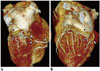
Song et al. (1) designated the subtypes R1, R2, L1, L2, and L3, and reported their frequency as 55%, 0%, 33.3%, 11.7%, and 0%, respectively. In a different study, the respective frequencies were 56%, 1.2%, 21.5%, 19.5%, and 0.4% (2). However, there is another subtype that was not mentioned in both studies. In this rare subtype, the sinoatrial node artery originates from the distal part of the right coronary artery (RCA) and courses along the posterolateral wall of the left atrium toward the groove between the left atrial appendage and left superior pulmonary vein (Fig. 1). The distal course of this artery is similar to the S-shaped sinoatrial node artery originating from the left circumflex artery. This rare subtype can be appropriately designated R3, consistent with the prior classification (1, 2). We retrospectively analyzed the coronary CT angiography results of 875 consecutive patients and found that only two cases (0.2%) showed this type of origin and course of the sinoatrial node artery. The frequency of RCA originating from the S-shaped sinoatrial node artery has been reported as 0.25% and 0.4% (3, 4). Since an unusual course of a coronary artery is important, especially in cardiovascular surgery, this rare subtype should be recognized and pointed out by radiologists.
 XML Download