

 PDF
PDF ePub
ePub Citation
 Print
Print


INTRODUCTION
Osteochondral lesions involve the articular cartilage and underlying subchondral bone, and most often affect the knee and the talus (1). Osteochondral lesions of the femoral head are uncommon, and few studies have reported the imaging findings of such lesions. Since this condition occurs in relatively young patients, and osteochondral damage puts joints at high risk of subsequent degeneration (2), timely recognition of this disorder is important for proper treatment. Herein, we review previous literature related to osteochondral lesions of the femoral head and 796report a case in which lesion involving the bilateral femoral heads was observed. This lesion manifested as subchondral cysts according to the initial radiography images, and led to further evaluation by computed tomography (CT) arthrography and immediately obtained magnetic resonance imaging (MRI) scan that revealed overlying articular cartilage defects. This report was approved by our Institutional Review Board.
CASE REPORT
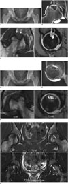
A 20-year-old female patient was referred to our hospital from a local hospital. Her chief complaint was pain in both hips along with severe right side pain of approximately 2 months duration. She was working in a restaurant and did not have any particular trauma history. Notably, however, she had been a taekwondo athlete in high school for several years. On clinical examination, both hips had full range of motion and revealed no crepitus or instability. Laboratory studies showed normal hematologic and biochemical values. Plain radiography of the hip showed well-defined subchondral radiolucent lesions with a subtle sclerotic rim in the superior portion of the right femoral head (Fig. 1A). This lesion was thought to be subchondral cysts. The acetabuli in both sides of the hip displayed mild dysplasia with lower limit of normal (center-edge angle, 24-25 degrees). No signs of degeneration, such as joint space narrowing or juxta-articular sclerosis, were observed, except for subtle osteophyte around the head-neck junction of the right femur. Deformations of the femoral head, such as flattening, or the presence of crescent signs were also absent. No sign of femoroacetabular impingement was noted. Based on imaging findings, a provisional diagnosis of avascular necrosis with subchondral cysts could not be excluded. To further investigate the lesion, CT arthrography and immediately obtained MRI scan of the right hip were performed. The respective images (Fig. 1B, C) revealed several subchondral cysts that were hypointense on the T1-weighted images (repetition time [TR]/echo time [TE], 374/20) and hyperintense on the T2-weighted fat-suppressed images (TR/TE, 1500/29). The overlying portion in this region revealed a focal articular cartilage defect measuring about 10 × 15 mm in size, consistent with an osteochondral lesion. No surrounding bone marrow edema was observed, and no cartilage damage was noted on the opposite site of the acetabulum. The scintigraphy scan with 99m technecium methylene diphosphonate showed no increased activity in the femoral head. On arthroscopy, chondral damage was noted in the superior portion of the femoral head. Thus, core decompression with bone graft harvested from the ilium was performed. Follow-up plain radiography of the hip performed about a year later revealed subchondral cysts in the superior portion of the left femoral head (Fig. 1D). This observation was similar to the lesion manifest in the right femoral head. The patient reported no incidence of trauma in the meantime, other than mild hip discomfort. CT arthrography (Fig. 1E) and immediately obtained MRI (Fig. 1F) scans of the left hip also showed several subchondral cysts, measuring about 14 × 13 mm in size, with associated overlying articular cartilage loss. This defect was also diagnosed as an osteochondral lesion of the left femoral head. Retrograde inspection of previous hip MRI scans performed at the local referral hospital 2 months before the first visit to our hospital revealed lesions of the left femoral head with abnormal signal intensity, including hypointensity on the T1-weighted images and hyperintensity on the T2-weighted images (Fig. 1G). This led us to hypothesize that the subchondral cyst lesion had progressed from a pre-existing lesion already present one year before. The chondral defect was confirmed by arthroscopy and core decompression with a bone graft was performed, as had been done for the right side.
DISCUSSION
Osteochondral lesions involve articular cartilage and the underlying subchondral bone, and mainly affect the knee and the talus (1). Osteochondral lesions of the femoral head are rare and the prevalence is unclear (3). Detailed reports of the imaging findings of such lesions are also quite limited. Therefore, osteochondral lesions of the femoral head can be easily overlooked, unless radiologists are suspicious of osteochondral lesions that develop in the femoral region. Furthermore, osteochondral lesions of the femoral head are known to cause joint degeneration (2). Thus, timely diagnosis and treatment are very important.
In our case, plain radiography images showed several subchondral cysts in both femoral heads. We diagnosed the osteochondral lesion by identifying the accompanying overlying articular cartilage defects by CT arthrography and MRI, which were performed for further evaluation. We excluded a diagnosis of avascular necrosis due to the absence of a double line sign or a crescent sign of the femoral head on MRI. Moreover, no signs indicative of primary osteoarthritis, such as joint space narrowing or juxta-articular sclerosis in both the acetabulum and femoral head, were observed.
The causes of osteochondral lesions have not yet been elucidated, and are likely to be multifactorial (4). One of the well-known causes of the osteochondral lesion of the femoral head is Legg-Calvé-Perthes disease. In this secondary lesion, however, separated subchondral bone with articular cartilage from the joint surface is the main feature, which is different than our case. Other etiologies of osteochondral lesions in the hip include previous infection, genetics, ischemia or trauma (5, 6). Many studies have reported damages incurred by repetitive traumas (7, 8, 9). Weaver et al. (10) reported normal plain radiographic finding but MRI detection of a subchondral lesion in an athlete with continuous hip pain, and suggested that although an athlete may not recall a specific incidence of past trauma, shearing injury of the cartilage by subluxation during joint rotation or impact damage caused by forces exerted by jumping could have occurred.
Kusumi et al. (7) demonstrated that osteochondritis dissecans of the elbow can result from articular cartilage damage inflicted by repeated trauma. Nakamura et al. (6) reported an osteochondral lesion of the femoral head believed to have originated from repeated impact and high axial loads in a fencer. Our patient was previously an athlete, and so could have developed the osteochondral lesions by repetitive trauma, despite the lack of recollection of any particular past trauma.
Weaver et al. (10) also reported abnormal bone marrow signals from an osteochondral lesion on the femoral head. This lesion exhibited hypointensity on the T1-weighted images and hyperintensity on the T2-weighted images, along with cartilage surface irregularities of the medial portion. In our case, although the cartilage surface irregularities were not immediately apparent from the previous MRI scans taken at the local referral hospital, lesions displaying hypointensity on the T1-weighted images and hyperintensity on the T2-weighted images were present in the left femoral head. An articular cartilage defect lesion accompanied by subchondral cysts was found about a year later, similar to the lesion previously observed on the right femoral head. We concluded that an osteochondral lesion was already present at the time of the previous MRI scan, and that this lesion progressed to the lesion with subchondral cysts.
It may often be necessary to distinguish osteochondral lesions from avascular necrosis in differential diagnoses. The articular cartilage of the femoral head has been shown to be unaffected in the majority of cases of avascular necrosis, whereas this cartilage is often damaged in osteochondral lesions. Moreover, avascular necrosis of the femoral head manifests itself as a somewhat broader lesion, often accompanied by serpiginous, low signal lines or crescent signs (11). On the other hand, osteochondral lesions often reveal smaller-sized areas with wedge-shaped signal abnormalities, or a narrow inlet with deep nest-shaped lesions, as seen in this case (12, 13). Patient age, in particular being younger, is also helpful in distinguishing this condition from avascular necrosis (10).
The signal changes of femoral heads reported by Weaver et al. (10) are thought to be indicative of lesions in the acute or subacute stage. In the chronic stage, however, a progressed degenerative bony lesion in the form of subchondral cysts, as seen in our case, or perhaps even a sclerotic change exhibiting hypointensity on the T1- and T2-weighted images, may be observed (6). Although some authors have reported that high uptake on the scintigraphy represents inflammation or lesion healing, no increased uptake was observed in our case, probably because the lesions are in the chronic stage.
Radiologists should bear in mind that osteochondral lesions can manifest themselves in various forms. In addition, young patients, especially those with a previous trauma or a history of being an athlete, are potential candidates for osteochondral lesions. Radiologists should look for bony lesions associated with articular cartilage defects in the femoral head, which could be indicative of an osteochondral lesion, similar to the lesions that are known to occur in the knee or the talus.
 XML Download