

 PDF
PDF ePub
ePub Citation
 Print
Print


INTRODUCTION
A chamber rupture is a rare type of cardiac injury caused by blunt chest trauma. The left ventricle (LV) is the most vulnerable site for chamber rupture, followed by the right ventricle (RV) (1). We report a rare case of RV pseudoaneurysm in a 53-year-old female who survived a 20-meter fall.
CASE REPORT
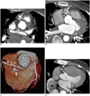
A 53-year-old woman was referred to our emergency room after a 20-meter fall. The patient presented a blood pressure of 45/35 mm Hg, pulse rate of 118/min and spontaneous breathing of 20/min. Her Glasgow Coma Score was 7. The patient underwent an emergency CT scan that revealed hemoperitoneum, hemopneumothorax and multiple fractures. A chest CT scan indicated an abnormal small round area of high attenuation adjacent to the RV (Fig. 1A). The patient underwent an emergency laparotomy to control hemorrhaging due to hemodynamic instability.
On the eighth day of admission, echocardiography was performed to evaluate cardiac function and identify RV pseudoaneurysm. The echocardiography failed to identify a pseudoaneurysm, and cardiac function was normal. Consequently, the patient underwent an electrocardiography (ECG)-gated cardiac CT scan for final diagnosis on the fifteenth day of admission. The ECG-gated cardiac CT scan demonstrated a 15 × 6 mm round high attenuation-collection (i.e., pseudoaneurysm) connected to the RV by the neck. On the twentieth day of admission, the patient underwent a follow-up CT scan to evaluate the stability of the aneurysm. On follow-up CT scan, the size of pseudoaneurysm had increased to 17 × 7 mm (Fig. 1B, C), but surgery was deferred due to the critical condition of the patient. The patient received medical treatment with close monitoring. Follow-up CT scan obtained one month later indicated interval decrease of the pseudoaneurysm. Final CT obtained two months later demonstrated spontaneous obliteration of the RV pseudoaneurysm (Fig. 1D).
DISCUSSION
Cardiac injury in patients with blunt chest trauma is estimated at 15% (1). The most common types of injury are chamber rupture, valve rupture and coronary artery rupture.
Several hypothesis explain the formation of post-traumatic myocardial pseudoaneurysm. Myocardial contusion leads to myocardial necrosis, rupture of the ventricle and pseudoaneurysm formation (2). Ischemic necrosis from vascular injury leads to pseudoaneurysm formation (3). Myocardial dissection, beginning as a small endocardial tear, leads to pseudoaneurysm formation (4). Consequently, the likely mechanism in this case was myocardial contusion or dissection, since the corresponding vessel was intact.
Left ventricular pseudoaneurysm occurs most frequently after myocardial infarction (5). Trauma accounts for only 7% of etiology. The clinical course of LV pseudoaneurysm, unlike true ventricular aneurysm, is relatively poor due to the high tendency of rupture and sudden death (6). Common symptoms and signs of LV pseudoaneurysm are heart failure, chest pain and dyspnea. Patients can also present nonspecific features such as cough, altered mental status and dizziness. The role of CT angiography is important, since 12% of patients with LV pseudoaneurysm can be asymptomatic (5).
Right ventricular perforation occurs after invasive procedures such as central venous catheterization, pacemaker implantation and endomyocardial biopsy (7). No report has specified the incidence of traumatic RV pseudoaneurysm after blunt chest trauma, and we estimate it is rare. Rupture is less common in RV pseudoaneurysm due to the lower pressure in the right-side circulatory system. Low pressure in the right side of the heart usually allows spontaneous obliteration of the neck. However, RV peak systolic pressure will increase if the patient has pulmonary hypertension with a subsequent increased risk of RV rupture (8). The patient did not have pulmonary hypertension, and the aneurysm resolved due to obliteration of the neck.
Shen et al. (9) reported on a real-time three-dimensional echocardiography that provided an excellent visualization of RV pseudoaneurysm. However, echocardiography failed to detect RV pseudoaneurysm despite a high suspicion of pseudoaneurysm based on CT. CT scan represents a more powerful diagnostic tool for patients with multiple trauma (10). An ECG-gated CT scan can provide more detailed anatomic information that indicates the presence (or absence) of pseudoaneurysm, size of pseudoaneurysm, neck, the origin of the pseudoaneurysm and myocardial enhancement pattern. In the acute or subacute phase, ventricular pseudoaneurysm looks like a round sac, located outside the ventricle, with high attenuation on the contrast enhancement scan. A narrow neck usually connects the pseudoaneurysm and the ventricle. In the chronic stage, this narrow neck is widened and the wall of the pseudoaneurysm may be calcified (11).
Surgical repair is the preferred treatment for LV pseudoaneurysm due to the high risk of rupture (12, 13, 14). However, RV pseudoaneurysm may be successfully managed with conservative treatment. Ahmad et al. (8) reported on nonoperative management with close monitoring that may be considered in the absence of tamponade, severe pulmonary hypertension, deterioration in hemodynamic status or serial imaging studies that indicates the expansion of the pseudoaneurysm. In this case, the size of the RV pseudoaneurysm increased on the first follow-up CT. We decided on conservative management instead of surgery due to the critical condition of the patient. The aneurysm decreased on follow-up CT obtained one month later and resolved on the follow-up CT obtained two months later without operation.
CONCLUSION
We described an unusual case of traumatic RV pseudoaneurysm in a patient after a 20-meter fall. We decided on conservative management despite the size increase indicated by short-term follow-up CT, due to the critical condition of the patient. The pseudoaneurysm spontaneously resolved without operation. Knowledge of imaging findings for RV pseudoaneurysm and its natural course may lead to proper diagnosis and management of the lesion.
 XML Download