

 PDF
PDF ePub
ePub Citation
 Print
Print


INTRODUCTION
Cardiac adenosine stress perfusion magnetic resonance (MR) imaging is a highly accurate diagnostic tool for coronary artery diseases (1-3). It measures the contrast enhancement of the myocardium during the first pass of a contrast agent bolus as it is sensitive to changes in the myocardial blood flow. During the imaging of a patient after a coronary artery bypass graft (CABG) surgery, the first pass kinetics of a contrast agent bolus are complex due to the altered distance of bypass graft vessels to the myocardial territory. Herein, we report about a case in which a delay in contrast agent arrival was observed at the myocardial territory whilst the patent bypass graft, mimicking a perfusion defect on adenosine stress perfusion MR images. The report also compares the semiquantitative perfusion parameters of this myocardial territory by the patent bypass graft with those of normal myocardium and of another myocardial territory with a stenotic bypass graft in the same patient.
CASE REPORT
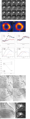
A 50-year-old man visited our hospital for a 3 month-follow-up examination after coronary bypass graft surgery. In this surgery a left internal thoracic artery (LITA) graft to the distal left anterior descending artery (LAD), and a right saphenous venous Y graft from the LITA to the diagonal branch and posterior descending artery (PDA) was performed through sequential anastomosis. The cardiac adenosine stress perfusion MR imaging revealed two perfusion defects at the mid-anterior and mid-inferior walls (Fig. 1A, Movies I, II). One perfusion defect at the mid-anterior wall was reversible on rest perfusion images. However, the other perfusion defect at the mid-inferior wallwas persistent on rest perfusion images. But later it disappeared on both stress and rest perfusion images. On the myocardial single-photon emission computed tomography, the inferior wall showed a mild degree of perfusion decrease which was considered as due to the diaphragmatic attenuation effect rather than an ischemic perfusion defect while the mid-anterior wall showed a reversible perfusion defect indicating a reversible ischemia (Fig. 1B). On cine images no regional wall motion abnormalities and on late gadolinium enhancement images no delayed myocardial enhancement were observed.
Signal time curve analyses at stress and rest perfusion MR were performed at three different regions: normal myocardium at the mid-lateral wall and two perfusion defects at the mid-anterior and mid-inferior walls (Fig. 1C). From the data, the perfusion index and ratio index of each region were calculated using a previously described method (4). In brief, a smooth-fit corrected signal-time curve within the time window of the first pass was analyzed using the gamma-variate function after subtraction of the baseline signal intensity (SI) value from the mean SI of each region. The time window of the first pass was determined from the SI curve of the left ventricular cavity. The time window of the first pass in the myocardium at the same level as the left ventricular cavity was determined by shifting the time window determined from the left ventricular cavity to when the SI in the myocardium started to rise. The maximal upslope indicating the perfusion index was computed from the peak value of the time derivatives of the fit function in the myocardial region, normalized by the maximal upslope in the left ventricular cavity. The ratio index, widely accepted to represent myocardial perfusion reserve, was defined as the ratio of maximal upslope at stress to that at rest (4). To calculate perfusion parameters an in-house software was developed using MATLAB 2012a (MathWorks, Natick, MA, USA).
The corrected signal-time curves demonstrated that the maximal upslope of the anterior wall decreased from 0.089 during rest to 0.022 during stress perfusion with a ratio index of 0.25, indicating typical reversible ischemia (Fig. 1D, E). In contrast, the maximal upslope of the mid-inferior wall was similar to that of normal myocardium during stress and rest perfusion. But the time to delivery and time to peak enhancement were delayed 7 seconds on both stress and rest images approximately (Fig. 1D). The maximal upslope of the mid-lateral wall increased with a ratio index by 1.10 from 0.094 during rest perfusion to 0.104 during stress perfusion. Also for the mid-inferior wall the maximal upslope increased with a ratio index by 1.10 from 0.079 during rest to 0.087 during stress perfusion (Fig. 1E). Coronary angiography revealed a focal tight stenosis at the anastomosis site between the right saphenous venous graft and diagonal branch, limiting the flow to the diagonal branch. With this can be explained the reversible ischemia at the apical to the mid-anterior wall on perfusion MRI. However, there was no significant stenosis at the right saphenous venous graft to the PDA. The contrast delivery in the right saphenous venous graft was slightly delayed compared to the LITA to distal LAD graft. It may indicate that the early perfusion defect at the mid-inferior wall was a result of delayed contrast media delivery due to the wide and long pathway of the bypass graft (Fig. 1F). Six-month follow-up images showed still a persistent perfusion defect without significant stenosis of the saphenous venous Y graft in the mid-inferior myocardial wall (Fig. 1G).
DISCUSSION
This case report illustrates focal delayed myocardial perfusion at the mid-inferior wall after CABG surgery mimicking a perfusion defect on adenosine stress perfusion MR images. There was a significant delay of contrast agent arrival (7 seconds) observed on the semiquantitative analysis of the perfusion at the mid-inferior wall myocardium. However, the maximal upslope of the corrected signal-time curves and rest-to-stress ratio index of maximal upslope were similar to those of normal myocardium. On coronary angiography, there was no significant stenosis at the bypass graft to the mid-inferior myocardial territory. Therefore, the early perfusion defect at the mid-inferior wall on stress and rest perfusion MRIs can be interpreted as a result of delayed delivery of contrast media due to the wide and long pathway of the venous graft.
Coronary artery bypass graft is a widely performed treatment option for coronary artery diseases. Since an exercise electrocardiogram is limited in many post-bypass patients, non-invasive stress imaging tests are often preferred to assess the patency of the graft (5). Although adenosine stress perfusion MR has been accepted as an accurate diagnostic tool, there have been a limited number of studies in post-bypass patients only with mixed results (6, 7). One study reported about reduced diagnostic accuracy in patients with CABG and attributed this to the different flow and perfusion kinetics involved (6). Another study reported about good diagnostic accuracy for the detection and localization of significant stenosis, but also about a reduced sensitivity compared with published data for patients without CABG (7). These results were obtained by visual assessment of myocardial perfusion.
In the present study, a semiquantitative analysis method was employed to better evaluate regional perfusion abnormalities. Among the semiquantitative parameters, upslope at stress is known to best detect ischemic myocardial segments and has been shown to correlate well with angiography and positron emission tomography (2, 8). In our case, maximal upslopes at stress in both the normal myocardium and myocardium perfused by the patent graft with a long pathway were higher than that of ischemic myocardium. Similar to a previous study the rest-to-stress ratio index of the maximal upslope of the inferior wall perfused by the patent graft with a long pathway was also similar to that of normal myocardium in the presented case (4, 9). Su et al. (4) reported that upslope significantly increased from rest to stress in normal myocardium, whereas in ischemic myocardium the change was insignificant. In another study, Kelle et al. (9) calculated the semiquantitative parameters of 38 patients after CABG surgery and reported similar wash-in kinetics (upslope) with a short delay in contrast arrival in areas perfused by bypasses compared to native coronaries. They observed that the mean contrast delay was below one beat in heartbeats and therefore concluded that differing contrast kinetics through graft vessels might not be a limiting factor in the accuracy of adenosine stress perfusion MR in post-CABG patients. Our case exhibited a contrast delay of 7 seconds which is much more delayed than in Kelle et al.'s (9) observation. The authors therefore postulate that the longer pathway of the graft and the size discrepancy between the graft and anastomotic native vessels may cause a greater delay in contrast delivery.
There are two concerns regarding our speculation. First, the delayed perfusion may be influenced by the collateral flow from the native coronary artery. However, we consider it unlikely as all native coronary arteries were completely occluded. Second, the PDA graft is a sequential Y graft of the saphenous vein and there is a tight stenosis at the proximal anastomotic site of diagonal branch. Thus, flow disturbances in the distal sequential graft due to proximal stenosis may have caused this perfusion defect more than the long graft course and size discrepancy. However, in this case, the tight stenosis was located in the proximal diagonal branch affecting the diagonal branch flow only. Interestingly, on follow-up coronary angiography taken after 6 months, the diagonal branch regressed due to tight stenosis and no stenosis was detected in the Y graft of saphenous vein. Follow-up perfusion MR images at both stress and rest after spontaneous regression of the diagonal branch still showed the same feature of a persistent perfusion defect in the inferior myocardial wall, indicating a delayed perfusion rather than a true perfusion defect. There was still neither delayed enhancement nor regional wall motion abnormality in that area. These imaging findings also support our speculation.
This case illustrates focal delayed perfusion after bypass surgery mimicking a perfusion defect on adenosine stress perfusion MR images. The delayed perfusion observed in our study also mimicked a perfusion defect in the myocardial SPECT study. However, the semiquantitative analysis of corrected signal-time curves revealed that the myocardial territory in question was not ischemic but presented a delayed perfusion due to the long pathway of the bypass graft.
 XML Download