Abstract
Calcifying aponeurotic fibroma is a rare, benign fibroblastic tumor. The lesion has a propensity for local invasion and a high recurrent rate. Therefore, accurate preoperative diagnosis and complete excision are important to prevent the recurrence of the tumor after surgical removal. However, radiographic and magnetic resonance imaging findings of calcifying aponeurotic fibroma have been extremely rarely described in the radiology literature. Thus, we report a rare case of calcifying aponeurotic fibroma affecting the dorsal wrist in a 67-year-old man, describe radiographic and MR findings, and discuss the differential diagnosis of the tumor.
Calcifying aponeurotic fibroma was first described and referred to as juvenile aponeurotic fibroma by Keasbey in 1953 (1). It is a rare, benign, locally aggressive fibroblastic soft tissue tumor that typically occurs in the palm of the hand and in the sole of the feet in children and adolescents (1-3). The lesion has a tendency to infiltrate the surrounding tissue. Extrinsic cortical erosion of the adjacent bone is rarely seen. The tumor has a predilection for local recurrence after surgical resection (2, 3). To our knowledge, there have been only few radiology studies describing the MR findings of calcifying aponeurotic fibroma. Furthermore, previous articles mainly investigated radiologically non-calcified masses in young children (3, 4). We report a very rare case of aponeurotic fibroma in a 67-year-old man with a 20-year clinical history that presented with a relatively densely calcified mass affecting his dorsal wrist. The purposes of this article are to report the radiographic and MR findings of calcifying aponeurotic fibroma and to provide useful information for the differential diagnosis of the lesion.
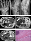
A 67-year-old healthy man presented with a 20-year history of a painless, slow growing mass in the dorsum of his right wrist. Physical examinations showed a firm, non-tender, palpable mass, measuring 1.5-cm, and overlying the carpal bones in the dorsum of his wrist. Radiographs demonstrated a relatively ill-defined soft tissue mass with speckled calcifications on the dorsal ulnar aspect of the wrist. Cortical scalloping of the underlying carpal bones was demonstrated (Fig. 1A-C). MRI showed an ill-defined rounded subcutaneous soft tissue mass partially encasing the tendons of the extensor carpi ulnaris (ECU) and the extensor digiti minimi (EDM) on the dorsum of the wrist. MRI revealed low to intermediate signal intensity on the T1-weighted image (T1WI) and heterogeneous mixtures of low and high signal intensity on the T2-weighted image (T2WI). The mass showed heterogeneous intense contrast enhancement after intravenous gadolinium administration. The lesion had a poorly demarcated margin and was associated with edema like signal changes in the adjacent soft tissues on MR images (Fig. 1D-F). In view of the radiographic and MR findings, the preoperative differential diagnosis included soft tissue chondroma, bizarre paraosteal osteochondromatous proliferation (BPOP), giant cell tumor (GCT) of the tendon sheath, fibroma of the tendon sheath, nodular fasciitis and a small sized synovial sarcoma. The mass was excised and was revealed to be an off-white colored, firm lesion. The mass adhered to and involved the tendon sheaths of ECU and EDM, the adjacent extensor retinaculum, and the wrist joint capsule with the intracapsular extension. The triquetral and hamate cortical bone erosions were seen. However, the mass could be excised without any resection of the bones or tendons. Microscopically, an ill-defined tumor mass showed multifocal calcifications surrounded by dense fibrous stroma. The fibrous stroma revealed mixed cellular areas near the calcification and lesser cellular fibromatosis-like areas in the background. Multilayers of fibroblasts with small round nuclei, histiocytes and multinucleated giant cells in the cellular areas were adjacent to the calcification. Lesser cellular and densely collagenous areas showed early chondoid metaplasia-like cellular changes such as small round nuclei and perinucler clear spaces in the hyalinized stroma. There were neither cellular atypism nor mitosis in the fibroblasts (Fig. 1G). A pathologic diagnosis of calcifying aponeurotic fibroma was made.
Calcifying aponeurotic fibroma is a rare, benign, locally aggressive fibroblastic tumor. The tumor usually appears in the first to second decade of life, although cases have been reported for ages ranging from birth to 67 years. Male patients are twice as commonly affected as female patients. Calcifying aponeurotic fibroma typically occurs in the distal extremities, most commonly in the fingers, palms and soles (1-3). However, this lesion has been observed at other sites, including the neck, mandible, forearm, elbow, knee and thigh (3). The tumor has been typically described as a firm, non-tender, slow growing mass, measuring less than 3 cm in diameter. It has a tendency to infiltrate the surrounding tissue, and has a predilection for local recurrence after surgical resection. The recurrent rate is approximately 50% (1-3). Calcifying aponeurotic fibroma, although locally recurrent, generally does not metastasize. However, there have been two reports of malignant transformation of calcifying aponeurotic fibroma. Lafferty et al. (5) made a pathologic confirmation of a juvenile aponeurotic fibroma that had metastasized to the lung and bone five years after its secondary excision. Enzinger and Weiss (6) stated that malignant transformation of aponeurotic fibroma was not found in the registry of the Armed Forces Institute of Pathology but that they had seen one case in consultation. Histologically, the lesion reveals characteristic scattered chondroid or calcific nodular foci surrounded by rounded, chondrocyte-like cells arranged in a linear or palisade pattern. There is less cellular, spindle fibroblastic component with a fascicular pattern between the coalescent calcified or chondroid nodules and emanating into the surrounding soft tissue. Mitotic figures are rare (7). According to a pathologic analysis of 22 cases by Fetsch and Miettinen (7), chondroid foci and mineralization were demonstrated in all but one case, regardless of radiographic evidence of calcification. Enzinger and Weiss (6) suggested the existence of biphasic development of the tumor, consisting of the initial and late phases. In the initial phase, which is seen more often in the young, the tumor has infiltrative and destructive growth and often lacks calcification. In the late phase, the tumor is more compact and nodular, and shows more prominent calcification and cartilage formation. Therefore, imaging appearances of calcifying aponeurotic fibroma can vary depending on the patient's age, presence of calcifications, and osseous involvement (6). Base on the literature, the mass seen in this case is considered to clinically and radiologically correspond to the late phase of calcifying aponeurotic fibroma, because the mass had a nodular appearance with relatively dense calcifications in our advanced aged patient with a long clinical history. Radiographic features of calcifying aponeurotic fibroma include an ill-defined or poorly demarcated margin of the tumor and a variable extent of fine, stippled calcifications within the lesion. Extrinsic erosion of the adjacent bone is very rarely seen (3). All these radiographic features with relatively dense calcifications were evident in our case (Fig. 1A-C). Computed tomography scan is optimal for depicting the calcified area of the lesion, with other lesions demonstrating non-specific soft tissue attenuation. MR imaging features of the mass have rarely been described in the radiology literature. Kwak et al. (4) reported a case of radiologically non-calcified calcifying aponeurotic fibroma in a young child, and described the MRI findings of the mass showing lower signal intensity than that of muscle on T1WI and T2WI, and intense heterogeneous enhancement after gadolinium administration. They stated that the low signal intensity on T2WI was attributed to the fibrous component and little cellularity of the mass. The dense calcifications of the mass in our case are considered to be also responsible for the low signal intensity on T1WI and T2WI, in addition to the fibrous component and little cellularity of the mass. When a small, slow growing, calcified or non-calcified soft tissue mass is identified in the wrist or hand, the differential diagnosis should include GCT of the tendon sheath, soft tissue chondroma, florid reactive periostitis or BPOP, fibroma of tendon sheath, and nodular fasciitis (8, 9). GCT of the tendon sheath is the second most common mass affecting the hand and wrist, after ganglion. The peak age is the third to fifth decade. Clinically, patients report asymptomatic, slowly growing masses attached to the tendon sheath and/or joint capsule. Radiographs may depict pressure erosion, and less commonly, periosteal reaction. There is no calcification within the lesion, unlike calcifying aponeurotic fibroma. MR images can optimally depict a well-defined mass with hemosiderin deposits. These findings may also be helpful to distinguish the lesion from calcifying apineurotic fibroma. Typical signal void artifacts can be seen on all sequences, particularly on gradient-echo images, and a more heterogeneous and predominantly low signal is found on T2WI. The lesion typically enhances after gadolinium administration (8). Soft tissue chondroma is a very rare well-defined cartilaginous mass. It has a predilection for the hand and wrist. Clinical presentation is of a slow growing mass of the hand or wrist with deep attachment to the tendon, tendon sheath, joint capsule, or periosteum. Radiographs may be negative, may demonstrate intralesional calcifications in 30% to 70% of cases, or may show extrinsic cortex erosion of the underlying bone (8, 9). MR imaging is highly specific and depicts a cartilaginous mass that demonstrates low signal intensity on T1WI and very high SI on T2WI. After contrast administration, small lesions show peripheral enhancement and larger lesions show more central enhancement. Intralesional low signal intensity may reflect calcifications (8, 9). Florid reactive periostitis and BPOP are considered to depict different phases of development of the same posttraumatic proliferative reaction (8). Florid reactive periostitis is also known as paraosteal fasciitis or periostitis ossificans (8). It usually involves the small bones of the hands and feet and usually occurs in young patients, between 20 and 30 years of age. Clinically, the patient usually has progressive painful swelling over the mass. The lesion is depicted as an ill-defined paraosseous mass with spare calcifications with florid lamellar or compact periosteal reaction on radiographs, which is an unusual feature for calcifying aponeurotic fibroma. The bone cortex is usually intact. BPOP also has a predilection for the small bone of the hand, and may widely occur in patients ranging from first to sixth decade. The patient typically has a painless mass. Radiograph shows a well-defined lobulated ossifying mass in the hand, usually less than 3 cm. MR imaging demonstrates low signal intensity on T1WI, varying high to low signal intensity on T2WI depending on the degree of its ossification (8). Fibroma of the tendon sheath presents as a slow growing, hard mass, firmly attached to the tendon. It shares imaging features and clinical presentations with GCT of the tendon sheath, but occurs much less commonly in the hand and wrist. MR signal is heterogeneous and non-specific. The lesion may appear as a well-defined, focal nodular mass with decreased signal intensity on all MR sequences and little or no contrast enhancement. MR signal intensity of the mass may vary depending on the degree of cellularity and myxoid component (8, 9). Nodular fasciitis is a non-neoplastic reactive lesion. The peak age is within the second to forth decade. It has a predilection for the volar aspect of the forearm. The hand and wrist are relatively uncommon sites. The mass may be mistaken for sarcoma due to its clinical presentation as a rapidly growing mass. Nodular fasciitis usually occurs in subcutaneous locations but can be intramuscular or fascial. Radiograph may demonstrate a soft tissue mass, which is rarely calcified (8, 9). MR imaging is non-specific, showing equal to or slightly higher signal intensity to muscle on T1WI, intermediate to hyperintensity on T2WI, and diffuse or peripheral enhancement after gadolinium administration. Features that may suggest the diagnosis of the lesion are linear extensions along the superficial fascia and mild surrounding edema (9). Malignant soft tissue tumor of the hand and wrist are extremely rare. Because they tend to be rather slowly growing, and are generally small at presentation, the lesion may be misdiagnosed as a benign tumor (8). Hence, calcifying aponeurotic fibroma should also be distinguished from the most common malignant soft tissue tumors in the hand and wrist, particularly in the presence of intratumoral calcifications, including epithelioid sarcoma, synovial sarcoma, and undifferentiated pleomorphic sarcoma (UPS) (8-10). Epitheliod sarcoma usually occurs in the flexor aspect of the upper extremity, and commonly affects young adults in the second to fourth decade of life. Radiograph occasionally shows calcifications in the tumor (9, 10). MR signal of the mass is non-specific. It may extend along the fascial planes with surrounding edema. There can be a prominent hemorrhage component resulting in a heterogeneous signal on T1WI and fluid-fluid levels. The mass usually contrast enhances (9, 10). Synovial sarcoma is derived rather from primitive mesenchymal cells than from synovial cells. The mass occurs in adults in their fourth decade. Two third of the tumor occurs in the lower extremities, particular near large joints. Approximately 30% of synovial sarcomas contain calcifications (9, 10). The tumor has relatively well-defined margins and can demonstrate fluid-fluid levels due to hemorrhage. MR signal of the tumor is mostly isointense to muscles with areas of high signal intensity consistent with hemorrhage on T1WI, and markedly heterogeneous signal intensity on T2WI (9, 10). UPS is a tumor formerly known as malignant fibrous histiocytoma, and is the most common type of soft tissue sarcoma in adults, but is not common in the hand and wrist (10). UPS typically presents as a painless, circumscribed, multinodular, lobulated tumor with degeneration. Calcifications or ossifications within the tumor can be seen in 5-20% of cases, usually curvilinear or punctuate, at the periphery of the lesion. Bone erosion or infiltration is common (10). MRI signal intensity is nonspecific, depending on the cellularity and myxoid content of the lesion and on the presence of hemorrhage, necrosis, and/or calcification. Extraskeletal chondrosarcoma and extraskeletal osteosarcoma are extremely rare in the hands and wrist. The main locations of extraskeletal myxoid chondrosarcoma are the deep soft tissues of the proximal extremities. The most common location of extraskeletal osteosarcoma is the thigh (10).
In summary, we report a rare case of relatively densely calcified calcifying aponeurotic fibroma in an elderly patient with radiographic and MRI features which have rarely have been described in the radiology literature. Because the recurrence rate of this tumor is high, accurate preoperative diagnosis and complete excision are important to prevent the recurrence of the tumor after surgical removal. Clinical, radiographic and MR imaging features of calcifying aponeurotic fibroma can be helpful in the preoperative differential diagnosis, and MR imaging is also helpful to evaluate the extension of the mass.
Figures and Tables
 | Fig. 1Calcifying aponeurotic fibroma of dorsal wrist in 67-year-old man.
AP (A), oblique (B) and lateral (C) radiographs of wrist demonstrate relatively ill-defined heterogeneously calcified soft tissue mass with extrinsic erosions (arrowheads in B) of adjacent carpal bones on dorsal ulnar aspect of wrist. Axial MR images (D-F) demonstrate ill-defined subcutaneous soft tissue mass, showing low to intermediate signal intensity on T1WI (D), heterogeneous mixture of low and high signal intensity on T2WI (E) and heterogeneously intense contrast enhancements on fat suppressed T1WI after intravenous gadolinium administration (F). Note that mass abuts tendons of extensor carpi ulnaris (short arrows in D-F) and extensor digiti minimi (arrows in D-F), erodes underlying carpal bones (arrowheads in D), and is associated with diffuse edema like signal changes in adjacent soft tissues of wrist. Photomicrograph (G) reveals scattered foci of calcification (white stars) and chondroid differentiation (asterisks) surrounded by peripheral less cellular, spindled fibroblastic component (arrows) between coalescent calcified and chondroid nodules (H&E stain, × 200). T1WI = T1-weighted image, T2WI = T2-weighted image
|
References
1. Keasbey LE. Juvenile aponeurotic fibroma (calcifying fibroma); a distinctive tumor arising in the palms and soles of young children. Cancer. 1953; 6:338–346.
2. Goldman RL. The cartilage analogue of fibromatosis (aponeurotic fibroma). Further observations based on 7 new cases. Cancer. 1970; 26:1325–1331.
3. Murphey MD, Ruble CM, Tyszko SM, Zbojniewicz AM, Potter BK, Miettinen M. From the archives of the AFIP: musculoskeletal fibromatoses: radiologic-pathologic correlation. Radiographics. 2009; 29:2143–2173.
4. Kwak HS, Lee SY, Kim JR, Lee KB. MR imaging of calcifying aponeurotic fibroma of the thigh. Pediatr Radiol. 2004; 34:438–440.
5. Lafferty KA, Nelson EL, Demuth RJ, Miller SH, Harrison MW. Juvenile aponeurotic fibroma with disseminated fibrosarcoma. J Hand Surg Am. 1986; 11:737–740.
6. Enzinger FM, Weiss SW. Fibrous tumors of infancy andchildhood. In : Enzinger FM, Weiss SW, editors. Soft tissue tumors. 3rd ed. St. Louis: Mosby;1995. p. 231–268.
7. Fetsch JF, Miettinen M. Calcifying aponeurotic fibroma: a clinicopathologic study of 22 cases arising in uncommon sites. Hum Pathol. 1998; 29:1504–1510.
8. Drape JL, Le Viet D. MR imaging of the fingers. In : Stoller DW, Li AE, Bredella MA, Potter HG, Rosenberg ZS, Bencardino JT, editors. Magnetic resonance imaging in orthopedics and sports medicine. 3rd ed. Philadelphia, PA: LW & W;2007. p. 1847–1932.
9. Sookur PA, Saifuddin A. Indeterminate soft-tissue tumors of the hand and wrist: a review based on a clinical series of 39 cases. Skeletal Radiol. 2011; 40:977–989.
10. van Vliet M, Kliffen M, Krestin GP, van Dijke CF. Soft tissue sarcomas at a glance: clinical, histological, and MR imaging features of malignant extremity soft tissue tumors. Eur Radiol. 2009; 19:1499–1511.


 PDF
PDF ePub
ePub Citation
 Print
Print


 XML Download