Abstract
Desmoplastic fibroma of bone is a rare locally aggressive, but non-metastatic tumor. In this case report, we present a desmoplastic fibroma in an unusual location, the clavicle. Desmoplastic fibroma involving the clavicle is extremely rare, with only 2 reported cases before 1985. We report the imaging findings of a desmoplastic fibroma of the clavicle with a review of the relevant literature.
Desmoplastic fibroma of bone is a rare locally aggressive but non-metastatic tumor which was first described by Jaffe in 1958 (1). The most commonly involved site is the mandible, followed by the femur and pelvis. In this case report, we present a desmoplastic fibroma in an unusual location, the clavicle. Bone tumors of the clavicle are rare, especially desmoplastic fibromas involving the clavicle are extremely rare, with only 2 cases reported in the literature before 1985 (2). We present here this unusual tumor with radiologic findings from conventional radiography, magnetic resonance imaging (MRI) and histopathology.
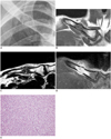
A 28-year-old man, with a history of sudden right shoulder pain during forceful flexion of his upper arm visited our hospital complaining of tenderness, resting pain, and limitation of range of motion due to pain. Initial work-up with plain radiograph demonstrated an oval shaped osteolytic lesion on the medial one-third of the right clavicle with pathologic fracture (Fig. 1A). Further evaluation with MRI was done after 10 days (Fig. 1B-D). The lesion showed hypointense to isointense signal compared to its adjacent muscle on T1-weighted images, and hypointense to isointense signal on T2-weighted images. After intravenous gadolinium contrast administration, the lesion presented heterogeneous enhancement. The mass showed a expansile bulging contour with endosteal erosion and periosteal reaction with enhancement. The aggressiveness of the tumor could not be evaluated easily due to the combined fracture. Our initial impression was benign primary bone tumor with low aggressiveness rather than malignant tumor. However, the patient was lost to follow up. Two years later, he came back to our hospital due to pain. Excisional biopsy was done. Histologically, the tumor was generally hypocellular to moderately cellular and composed of bland fibroblasts in a background of numerous thick and wavy collagen fibers. Thin spicules of residual trabecular bone with reactive changes were presented within the tumor. Multiple sections of the tumor presented no evidence of cellular pleomorphism, changes in the nuclear cytoplasmic ratio, nuclear hyperchromasia, or mitotic activity. Despite its bland appearance, the tumor showed multifocal soft tissue extension (Fig. 1E). These findings resembled those of soft tissue desmoids. Thus, the tumor was diagnosed as a desmoplastic fibroma of bone, which is considered as an intraosseous counterpart of soft tissue desmoids. Finally, a wide resection including the lesion and adjacent tissues was done with an allogenous fibular bone graft and an autogenous corticocancellous iliac bone graft.
Desmoplastic fibroma is a rare benign primary bone tumor, which histologically similar to the soft tissue desmoid tumor. The incidence of desmoplastic fibroma is reported to comprise 0.1-0.3% of all primary bone tumors. The mandible (22%), pelvic bones (13%) and long bones such as the femur (15%), radius (12%), tibia (9%) are known as the most frequent sites of involvement (2, 3). On the other hand, the clavicle is the only long bone with a transverse axis. Only 0.45-1.01% of all bone tumors are found in the clavicle. Furthermore, the majority of tumors, such as myeloma, osteosarcoma, and Ewing's sarcoma, are malignant (4, 5). Therefore, desmoplastic fibromas involving the clavicle are extremely rare, with only 2 cases reported in the literature before 1985 (2).
Clinical signs and symptoms are usually nonspecific. Pain and swelling are predominant symptoms, but some patients may be asymptomatic. Pathologic fractures are reported in 9-15% of cases (6-8).
In the literature, the most consistent radiographic findings are well defined non-sclerotic marginated geographic lesions (94%), with internal pseudotrabeculation (91%) and bone expansion (89%) (7, 8). These findings are also presented in our case. On MR imaging, histologically fibrous tissues are expected to show T2 shortening, and the majority of reported cases have shown hypointense or isointense signal on T1-weighted images, compared to the adjacent muscle, as well as hypointense or isointense signal on T2-weighted images. After intravenous gadolinium contrast administration, the mass shows a heterogeneous enhancement pattern, and these enhancement patterns seem to reflect the variable composition of the cellular part and collagenous matrix (9, 10). In our case, the mass showed hypointense to isointense signal on both T1 and T2 weighted images with a heterogeneous enhancement pattern, and showed enhancement of the adjacent bone marrow and periosteal soft tissue beyond the cortex.
On plain radiograph, this well-defined non-sclerotic marginated geographic lesion cannot be differentiated easily from various benign and malignant tumors. However, on MRI, radiologic features of a predominantly osteolytic lesion with prominent T2 shortening makes the diagnosis of demoplastic fibroma plausible (9). This T2 shortening is a differential feature from common clavicle tumors such as myeloma, osteosarcoma, and Ewing's sarcoma. But, some rare clavicle tumors such as non-ossifying fibroma, fibrous dysplasia, giant cell tumor, low-grade fibrosarcoma, and malignant fibrous histiocytoma also could show T2 shortening, so they needed to be included in differential diagnosis. Generally, non-ossifying fibroma and giant cell tumor show hyperintense signal on T2-weighted image. Non-ossifying fibroma and fibrous dysplasia usually show sclerotic borders on plain radiograph. Low-grade fibrosarcoma and malignant fibrous histiocytoma mostly show ill-defined border and soft tissue extension.
In summary, we report a desmoplastic fibroma of the clavicle presented with pathologic fracture. Desmoplastic fibroma should be considered in the differential diagnosis of a locally aggressive lesion with hypointense signal on T2-weighted images, although its involvement of the clavicle is very rare.
Figures and Tables
Fig. 1
Desmoplastic fibroma of clavicle in 28-year-old man.
A. Part of plain anteroposterior radiograph of right clavicle reveals well-defined non-sclerotic marginated geographic lesion on medial one-third of right clavicle, with pathologic fracture. B-D. Coronal (B) T1-weighted magnetic resonance image shows hypointense to isointense signal compared to adjacent muscle, and lesion also demonstrates hypointense to isointense signal on axial (C) T2-weighted image. Gadolinium enhanced T1-weighted coronal (D) image reveals heterogeneous enhancement of mass. Also, adjacent bone marrow and soft tissue along periosteum are enhanced. E. Microscopic examination shows bland fibroblasts in background of numerous thick and wavy collagen fibers without evidence of cellular pleomorhpism, changes in nuclear-cytoplasmic ratio or mitotic activity (H&E, × 200).

References
1. Jaffe HL. Tumors and tumorous conditions of the bones and joints. Philadelphia, PA: Lea and Febiger;1958. p. 298–303.
2. Gebhardt MC, Campbell CJ, Schiller AL, Mankin HJ. Desmoplastic fibroma of bone. A report of eight cases and review of the literature. J Bone Joint Surg Am. 1985; 67:732–747.
3. Böhm P, Kröber S, Greschniok A, Laniado M, Kaiserling E. Desmoplastic fibroma of the bone. A report of two patients, review of the literature, and therapeutic implications. Cancer. 1996; 78:1011–1023.
4. Smith J, Yuppa F, Watson RC. Primary tumors and tumor-like lesions of the clavicle. Skeletal Radiol. 1988; 17:235–246.
5. Pratt GF, Dahlin DC, Ghormley RK. Tumors of the scapula and clavicle. Surg Gynecol Obstet. 1958; 106:536–544.
6. Inwards CY, Unni KK, Beabout JW, Sim FH. Desmoplastic fibroma of bone. Cancer. 1991; 68:1978–1983.
7. Crim JR, Gold RH, Mirra JM, Eckardt JJ, Bassett LW. Desmoplastic fibroma of bone: radiographic analysis. Radiology. 1989; 172:827–832.
8. Taconis WK, Schütte HE, van der Heul RO. Desmoplastic fibroma of bone: a report of 18 cases. Skeletal Radiol. 1994; 23:283–288.
9. Frick MA, Sundaram M, Unni KK, Inwards CY, Fabbri N, Trentani F, et al. Imaging findings in desmoplastic fibroma of bone: distinctive T2 characteristics. AJR Am J Roentgenol. 2005; 184:1762–1767.
10. Vanhoenacker FM, Hauben E, De Beuckeleer LH, Willemen D, Van Marck E, De Schepper AM. Desmoplastic fibroma of bone: MRI features. Skeletal Radiol. 2000; 29:171–175.


 PDF
PDF ePub
ePub Citation
 Print
Print


 XML Download