

 PDF
PDF ePub
ePub Citation
 Print
Print


INTRODUCTION
Uterine artery embolization (UAE) is a safe and effective procedure for the control of postpartum hemorrhage (1). Detailed knowledge of uterine vascularization and ovarian artery supply to the uterus is necessary for the management of postpartum hemorrhage. The ovarian arteries usually arise from the anterior circumference of the abdominal aorta at the level of the renal arteries (2). Anatomic variations of the ovarian arteries are relatively common (2), and awareness regarding these variants is essential for successful UAE. Few studies have reported the abnormal origin or course of ovarian arteries (2, 3). Here, we present a very rare case of an aberrant ovarian artery arising from the common iliac artery.
CASE REPORT
A 46-year-old pregnant woman at 31 weeks gestation was referred to the gynecology department of our hospital for premature membrane rupture with uterine atony and placenta accreta. She had no significant past medical history, except a previous caesarean section. After an emergency caesarean section, the patient presented severe postpartum hemorrhage secondary to uterine atony and was referred to our department (interventional radiology).
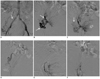
Embolization was performed using the single femoral artery approach with insertion of a 5-Fr introducer sheath (Terumo, Tokyo, Japan). At the beginning of the procedure, flush aortography (Fig. 1A) with a 5-Fr pigtail catheter (Cook, Bloomington, IN) positioned at the level of the celiac trunk showed a single aberrant artery arising from the right common iliac artery. Selective angiography of the aberrant artery using a 5-Fr Yashiro catheter (Terumo, Tokyo, Japan) revealed that it had a sinuous course and was tortuous (Fig. 1B). This artery showed the typical ovarian blush and abundant collateral perfusion to the markedly enlarged uterine fundus (Fig. 1C).
Both ovarian arteries could not be observed on pelvic aortography, and anastomosis with the uterine artery was not clearly defined. The diameter of the aberrant artery was larger than the diameters of both uterine arteries. Selective angiography of both the internal iliac arteries and both uterine arteries showed markedly increased uterus vascularity. The aberrant ovarian artery (Fig. 1D) and both uterine arteries (Fig. 1E, F) were embolized with 1 × 1 mm gelfoam pledgets (Johnson & Johnson, Skipton, UK) by using a 2.7-Fr microcatheter (Progreat®, Terumo, Tokyo, Japan). Vaginal bleeding ceased after this successful embolization procedure.
Three days after embolization, the patient underwent a contrast-enhanced abdominal-pelvic computed tomography (CT) scan, and no associated unusual findings such as anomalies of the kidney or renal arteries were noted.
The patient's condition rapidly stabilized after the embolization procedure, and she was discharged in a good condition.
DISCUSSION
Anatomic variations in the origin of ovarian arteries are relatively common (2), and awareness regarding these variants is essential to ensure successful UAE. Few cases of an aberrant ovarian artery originating from the inferior mesenteric artery or renal artery have been reported (2, 3). In the present report, we described an aberrant artery originating from the common iliac artery.
We suggested that this aberrant artery was an ovarian artery because of its characteristic sinuous course and tortuous shape. This artery supplied the right adnexa and a large portion of the enlarged uterine fundus. Its diameter was larger than the diameters of both uterine arteries. We believe that the enlargement of the aberrant ovarian artery was a response to pregnancy.
One of the reasons underlying embolization failure is non-embolized aberrant branches continuing to perfuse the uterus (4-6). If vaginal bleeding persists after UAE, aberrant arteries or collateral blood supply must be checked for and these arteries should also be embolized.
In conclusion, we described a very rare case of an aberrant ovarian artery arising from the common iliac artery supplying a large portion of the uterus. Currently, ovarian artery embolization is frequently performed, ever since the advent of UAE for treating postpartum hemorrhage. Therefore, precise knowledge of the anatomic variations of the ovarian artery is important for embolization to be successful.
 XML Download