

 PDF
PDF ePub
ePub Citation
 Print
Print


INTRODUCTION
Isolated left ventricular (LV) apical hypoplasia is a recently described congenital cardiac anomaly. Most reported cases of isolated LV apical hypoplasia have not been accompanied by other abnormalities (1-13). We herein present a case of a 33-year-old male patient with isolated LV apical hypoplasia combined with infundibular pulmonary stenosis and aortic stenosis. To our knowledge, this rare combination has not been reported in the English literature.
CASE REPORT
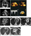
A 33-year-old male patient was referred to our hospital for evaluation of a cardiac murmur. The cardiac murmur was found incidentally during a preoperative evaluation for hemorrhoids. He had no history of congenital heart disease, and there was no family history of premature coronary artery disease, cardiomyopathy or sudden cardiac death. A continuous cardiac murmur was heard during the entire heartbeat. A chest radiograph showed no abnormalities. An electrocardiogram (ECG) indicated a normal sinus rhythm, right axis deviation, incomplete right bundle branch block, right atrial enlargement and right ventricular (RV) hypertrophy. Transesophageal and transthoracic echocardiography showed a spherically shaped LV and dilation of all cardiac chambers with good global LV systolic function (Fig. 1A). They also revealed heavily calcified aortic valve with moderate to severe stenosis (Fig. 1B), mild mitral regurgitation and RV outflow tract acceleration due to infundibular hypertrophy (Fig. 1C). Grade II diastolic dysfunction was also noted (E/A ratio, 1.9; isovolumic relaxation, 90 ms; deceleration time, 210 ms).
Cardiac magnetic resonance imaging (MRI) was performed using a 1.5-T scanner with standard ECG-based referencing (Siemens Symphony, Erlangen, Germany), and the findings supported and better defined the echocardiographic results. The MRI sequences were as follows: Anatomical evaluation was performed with transverse dark-blood HASTE (repetition time [TR]/echo time [TE], 700/27; matrix size, 256 × 123; slice thickness, 8 mm) with or without fat saturation. Dynamic evaluation was done with an echo-planar cine true fast imaging with steady-state precession (TR/TE, 66.2/1.3; matrix size, 192 × 113; slice thickness, 5 mm) in two-, three-, four-chamber and short axis views. Phase-sensitive inversion recovery turbo FLASH pulse sequence was also performed for the detection of LV myocardial scar (TR/TE, 826.4/3.3; matrix, 256 × 156; slice thickness, 6 mm) 10 minutes after intravenous injection of gadobutrol (0.2 mmol/kg). The LV exhibited a truncated and spherical configuration with rightward bulging of the interventricular septum and elongation of the RV. The papillary muscles originated from the flattened anterior apex (Fig. 1D). Quantitative RV ejection fraction was normal (52%). Transverse HASTE images outlined fat within the myocardium at the LV apex (Fig. 1E, F). There was no delayed enhancement of the myocardium to indicate myocardial fibrosis. Further, there was also infundibular pulmonary stenosis and aortic stenosis (Fig. 1G, H) without an atretic mitral valve. These echocardiographic and cardiac MRI anatomic features were consistent with isolated LV apical hypoplasia combined with infundibular pulmonary stenosis and aortic stenosis.
DISCUSSION
Isolated LV apical hypoplasia was first described in 2004 in a case report by Fernandez-Valls et al. (1). To our knowledge, there are 13 case reports involving 16 patients (mean age, 30.5 years; age range, 3 months to 63 years; six males) with this entity (1-13). Most of the reported cases of isolated LV apical hypoplasia were not accompanied by other cardiac abnormalities, with the exception of two cases. In those cases, the accompanying abnormalities were congenital patent duct arteriosus and complete transposition of the atrioventricular valves (2, 3). Our patient had infundibular pulmonary stenosis and aortic stenosis. To our knowledge, this is the first case of LV apical hypoplasia combined with infundibular pulmonary stenosis and aortic stenosis.
The pathogenesis of LV apical hypoplasia is unknown; however, the proposed mechanism is an abnormal ventricular septation during fetal cardiac development. Defective LV apical development may be due to relatively inadequate LV to RV dilatation during ventricular partitioning in the fifth week, resulting in a spherical LV with elongated RV wrapping (1).
Fernandez-Valls et al. (1) described the MRI and multidetector computed tomography findings of isolated LV apical hypoplasia as follows: 1) a truncated and spherical LV configuration with abnormal diastolic or systolic function, with exaggerated rightward bulging of the interventricular septum during diastole; 2) replacement of the LV apical myocardium, particularly along the distal interventricular septum, with fatty material contiguous with epicardial fat, suggesting deficient apical development; 3) anteroapical origin of the papillary muscle; and 4) elongation of the normally functioning RV wrapping around the deficient LV apex. Isolated LV apical hypoplasia should be differentiated from hypoplastic left heart syndrome, which is characterized by underdevelopment of the aorta, aortic valve and entire LV secondary to interrupted growth of the developing LV (1). Our patient had mature mitral valves, and the ventricular malformation was limited to the LV apex; hypoplastic left heart syndrome is associated with an atretic or stenotic mitral valve in 95% of cases (4, 5).
The reported cases revealed the differences in the clinical presentation among the age groups. Patients diagnosed during adulthood usually had symptoms (10/12, 83%) ranging from fatigue to exertional dyspnea, congestive heart failure with systolic or diastolic dysfunction, pulmonary edema and hypertension, or even malignant tachyarrhythmia. However, patients diagnosed during childhood were usually asymptomatic (4/4, 100%) (1-13).
The hemodynamics of isolated LV apical hypoplasia is similar to restrictive LV cardiomyopathy. In severe case, it results in biventricular failure, pulmonary hypertension and atrial or ventricular tachyarrhythmias (6). Most of the cases (11/16, 68%) described LV systolic and/or diastolic dysfunctions with decreased LV ejection fraction (1, 2, 6-10) or low normal value of LV systolic function (11).
Our patient has mild mitral regurgitation. Mitral regurgitation on echocardiography can be shown (5/16, 31%) due to the abnormal origin of a complex papillary muscle network. However, significant regurgitation was not common (1/16, 6%).
Although little is known about this entity, a close follow-up is required even in asymptomatic patients to monitor for signs and symptoms of heart failure, pulmonary hypertension and potentially malignant tachyarrhythmia (7, 9). Almost all of the reported patients responded well to the standard heart failure treatment with the exception of one, who died of ventricular fibrillation and subsequent hemodynamic deterioration (6).
Isolated LV apical hypoplasia is a recently recognized congenital cardiac anomaly that has a characteristic MRI appearance. We herein presented a patient with combined infundibular pulmonary stenosis and aortic stenosis. Our patient is currently on a regular expectant follow-up, with particular attention paid to the development of heart failure and tachyarrhythmias.
 XML Download