

 PDF
PDF ePub
ePub Citation
 Print
Print


INTRODUCTION
Dynamic contrast-enhanced magnetic resonance imaging (DCE-MRI) is an important diagnosis tool of breast disease because of its good soft tissue resolution and time-signal intensity curve; it has a higher sensitivity and often presents malignant breast nodules which are missed by physical examination, mammography and ultrasound (1-3). We present an interesting case of an asymptomatic 30-year-old female Asian patient with a small suspicious breast nodule which was not detected by breast palpation, mammography, and color Doppler ultrasound, but was detected by DCE-MRI. This nodule was surgically removed and pathologically confirmed as hemangioma, a kind of benign vascular tumor which is rarely seen in the breast.
CASE REPORT
We present an asymptomatic 30-year-old female Asian patient who gave birth 2 years ago. There is no history of hormone intake or family history of breast cancer. Ten months ago, the patient underwent breast palpation, mammography, and ultrasound (for the first time). The results present a nodule (1.6 × 1.2 × 0.9 cm3) locating on the right breast at 7 o'clock position with an oval shape, clear boundary, internal hypoechoic, and calcifications. Radiologists classify the lesion as BIRADS category 3. The patient requested the lesion to be surgically removed. This lesion was pathologically confirmed as breast fibroadenoma with calcification.
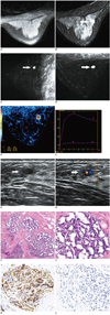
Before the surgery, MR scanning (GE Signa HDxt 3.0T) was performed according to the recommendation of a clinician. DCE-MRI did not detect enhancement of the mass (breast fibroadenoma with calcification). Surprisingly, a new small nodule (0.7 × 0.5 × 0.4 cm3) was found at the upper outer quadrant of the right breast. MRI presented an oval mass located on the mammary gland, anterior to the superficial pectoralis fascia with a well-defined margin. An unenhanced T1-weighted image did not display the mass well (Fig. 1A), while a T2-weighted image showed the mass area of high signal (Fig. 1B). DCE-MRI presented a strong homogeneous enhancement of the mass (Fig. 1C, D). The region of interest was obtained from the enhancing area of the lesion to generate a time-signal intensity curve. The curve was classified as washout type (Fig. 1E, F). Radiologists classify this mass as BI-RADS category 4B. Ultrasound was then carefully performed on the patient (for the second time), showing an oval nodule with a well-defined margin and internal hypoechoic (Fig. 1G). Color Doppler flow imaging presented rich blood flow signals (Fig. 1H).
This suspicious breast lesion was strongly advised to be surgically removed. Before surgery, two experienced radiologists made a surface marker for the lesion based on the DCE-MRI and ultrasound presentation. In the operating room, the surgeon confirmed the lesion again by ultrasound (for the third time) before the surgery. The lesion was pathologically confirmed as breast capillary hemangioma, which is a benign vascular tumor and rarely seen in the breast (Fig. 1I, J). Immunohistochemical staining showed strong positive CD34 expression and negative CK expression of the lesion, which supported our diagnosis (Fig. 1K, L).
DISCUSSION
Ultrasound is a very useful tool for checking breast diseases, especially in young women with mammary gland hyperplasia. While it has been reported that hemangiomas always show an oval or lobular shape with well circumscribed or microlobulated margins on sonography (4) (which were also seen in our case), these characteristics are not sufficiently specific. While hemangiomas often locate in a superficial position (subdermal or in the subcutaneous tissues) (5, 6) (which is similar to our case), this location makes it difficult to visualize the mass on regular craniocaudal and mediolateral oblique views, tangential views are necessary to prove the superficial nature of the mass.
Hypervascularity with multiple peripheral poles or internal vessels has been reported in malignant lesions. However, benign hemangiomas in the breast have also been described as displaying high vascularity.
MRI and DCE-MRI are frequently used to check for breast disease in Asian women because of the common occurrence of mammary gland hyperplasia and smaller size of female breasts. It is well known that MRI could detect inconspicuous lesions because of its good soft tissue resolution (7). Breast hemangiomas have been reported to show homogeneous hyperintensity on T2-weighted images as in our case. In a Gd-enhanced dynamic study, Vorela AL et al. reported that hemangiomas present early intensive enhancement followed by a plateau (8), which differs from our case (washout type curve, indicating malignant breast lesion), and the images and curve render it difficult to confirm the diagnosis. The difference of vascular morphology and numbers might be the main reason for the different DCE-MRI presentation and curve type.
Breast tumors contain angiosarcomas and hemangiomas (9). Angiosarcoma is rare but the most malignant of all breast neoplasm, with a mean survival time of about two years. This tumor presents as an ill-defined, unencapsulated, soft, spongy mass with dilated vascular channels. Hemorrhage and necrosis are common, especially in larger tumors. Histologically, the endothelial cells of angiosarcoma are usually large, plump, and single layered. Cellular atypia may be mild and mitoses are variable in number. Hemangiomas are benign vascular tumors of two common types (capillary or cavernous) that are based on the size of the vessels involved. In this case, microscope imaging showed that the hyperplastic vascular tumor has a regular morphology and clear boundary, has no interstitial infiltrates, no vascular branching structure, and the papilla structure could be found in the lumens. Endothelial cell atypia, papillary tufting, mitotic figures, necrosis, or other features of atypical vascular lesion or angiosarcoma were not identified. These findings were diagnostic of a hemangioma, predominantly capillary in pattern.
It has been reported that breast hemangioma could be found in 11% of postmortem specimens of the female (10). Nowdays, DCE-MRI is increasingly used to check breast diseases. If the DCE-MRI presents a small mass (usually ≤ 2 cm3) with a clear boundary, homogeneous enhancement, superficial location, and even the mass presents a washout type curve, alerting the radiologists that hemangioma could be a possible diagnosis.
 XML Download