

 PDF
PDF ePub
ePub Citation
 Print
Print


INTRODUCTION
Neurofibromatosis type 1 (NF1), or Von Recklinghausen disease, belongs to a group of heterogeneous diseases. It is of autosomal dominant inheritance and is caused by an abnormality of the long arm of chromosome 17 (1, 2). Many abnormalities are used to diagnose this disease according to diagnostic criteria, including: cafe-au-lait spots, plexiform neurofibroma, other forms of neurofibroma, Lisch nodules, fleckling, optic nerve glioma, and abnormal bony lesions; the disease presents in first degree relatives. This disease is associated with multiorgan involvement, primarily the central nervous system. However, vascular involvement, such as occlusion, stenosis, aneurysm or ectasia, is rare (2). We report a case of a spontaneous rupture of a left 5th intercostal artery aneurysm, presenting with massive left hemothorax; this case was successfully treated by transarterial coil embolization.
CASE REPORT
A 46-year-old Thai female presented with acute dyspnea and pleuritic chest pain which she had felt for one day. On general physical examination, she had tachypnea and subcostal retraction. Additionally, she had multiple café-au-lait spots on her trunk, plexiform neurifibroma on her left leg, severe kyphoscoliosis and pectus carinatum. Her blood pressure was 150/90 mm Hg, heart rate 34 beats/minute, and respiratory rate 122 breaths/minute. Oxygen saturation at room air was 85% and increased to 99% after she was given an oxygen mask with a bag 10 liters/minute. A chest examination showed decreased breathing sounds as well as dullness, on percussion, at the left lung; the trachea had shifted to the right. Her hematocrit value and hemoglobin levels were 30.6% and 9.4 mg/dL, respectively. Platelet count and coagulogram were within the normal limits.
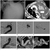
The patien's chest film revealed haziness in her left lung, mediastinum shift to the right, and severe kyphoscoliosis (Fig. 1A). She was referred to our department for an emergency computed tomography (CT) angiography of the chest in order to rule out a pulmonary embolism. The chest CT angiography did not reveal a pulmonary embolism. There was a massive left hemothorax with a total collapse of the left lung. An 8.0-mm saccular aneurysm was detected in the proximal left 5th intercostal artery, with adjacent sentinel hematoma (Fig. 1B). A ruptured aneurysm with massive left hemothorax was diagnosed. An incidental intrathecal cyst from T4-T8 levels, possibly thoracic meningocele, was also present (not shown). The patient was scheduled for an emergency embolization.
At the interventional unit, a chest tube was inserted, in order to relieve dyspnea, using a 10 Fr Pigtal catheter (Flexima, Boston-Scientific, Cork, Ireland). A large amount of flesh blood was obtained. Then, a digital subtraction aortogram was performed, using a 5 Fr Pigtail catheter (Boston-Scientific, Cork, Ireland) from the right common femoral arterial approach. Findings showed a small saccular aneurysm at the proximal left 5th intercostal artery, with adjacent distal arteriovenous fistula between the left 5th intercostal artery and vein, which drained into the hemiazygose vein (Fig. 1C). No contrast extravasation was detected. Selection of the left 5th intercostal artery was performed using a 5 Fr Mik catheter (Boston-Scientific, Cork, Ireland). Then, 5 mL of non-ionic contrast was administrated to clearly define the lesion and identify the proximal neck (Fig. 1D). A 2.7 Fr microcatheter (Progreat, Terumo, Leuven, Belgium) was advanced through the 5 Fr Mik catheter into the artery directly proximal to aneurysm. Following this, the embolization was performed via a microcatheter, using a single fiber platinum microcoil (4 mm-2 cm, Diamond-18, Boston-Scientific, Cork, Ireland).
The post-embolization angiogram showed successful occlusion of the aneurysm and arteriovenous fistula (Fig. 1E). Completion aortography confirmed complete occlusion of the lesion and an absence of other potential bleeding (Fig. 1F). After embolization, the patient's clinical signs improved. The chest tube was removed on the seventh day after embolization. Follow-up chest radiograph revealed near-complete resolution of left pleural effusion, and norecurrent bleeding occurred. The patient was discharged on the twelfth day after embolization.
At her oneyear follow-up, the patient's general condition was good, and CT angiography of the chest revealed complete occlusion of the prior aneurysm. However, there was recurrence of a small arteriovenous fistula (Fig. 1G, H). The re-embolization was recommended, but the patient refused the procedure because of no symptoms related to the fistula.
DISCUSSION
Neurofibromatosis type 1 is a disease that presents with multisystem involvement. The incidence of arterial involvement in this disease is rare:about 3.6% (1, 2), and is mostly in the form of stenosis or an aneurysm. A ruptured artery, arteriovenous fistula, or occlusion is also reported.
The cause of an arterial lesion in NF1 is considered by wall fragility. There are multiple studies describing the cause of an arterial lesion. Greene et al. (3) divided arterial lesions into two categories. In the first category, a large artery, such as the aorta, carotid, subclavain, and proximal renal artery is surrounded by neurofibromatous or ganglioneuromatous tissue resulting in intimal proliferation, thinning of media, and fragmentation of the elastic layer leading which leads to stenosis or aneurysm formation. In the second category, the smaller vessels are not related to neural malformation but to dysplasia of vessels. Leier et al. (4) described two causes of arterial rupture. Firstly, neurofibromatosis invades the muscular layer, which may reduce the strengthening of the vessel wall. Secondly, the vasa vasorum of a large vessel may be compressed by neurofibromatous tissue, resulting in a weakened segment of the artery secondary to ischemia. Reubi et al. (5) described the cause of vasculopathy related to NF1 in small vessels, including intimal proliferation, as resulting in luminal obstruction, intimal fibrohyaline thickening, thinning of media, elastic fragmentation and aneurismal dilatation.
We found, in the existing literature, only 27 cases of spontaneous massive intrathoracic hemorrhage in patients with NF (1, 2, 7). The largest series were 12 cases, reported by Miura and colleagues, in Japan (6). There were only 2 cases presenting with hemomediastinum, and the remaining cases presented as hemothorax.
The most common locations of arterial bleeding are the subclavain and intercostal arteries. However, bleeding from the thyrocervical trunk, internal thoracic artery, phrenic artery, and left vertebral artery were also reported.
Computed tomography angiography is the best modality for diagnosis. Moreover, it can be helpful in planning treatment that identifies the location of arterial bleeding prior to endovascular or surgical treatment. After treatment, CT angiography is used as it is readily available as a non-invasive procedure (8). Inthis case, the aneurysmally dilated intercostal artery remained patent even after coil embolization (Fig. 1E), which means there is a risk of re-bleeding. Therefore, to ensure long-term benefit to the procedure, follow-up (imaging) results are essential. For this patient, 1-year follow-up CT angiography revealed recurrence of a small arteriovenous fistula (Fig. 1G, H).
Spontaneous massive hemothorax is an emergency, life-threatening condition. In the past, surgery was the treatment of choice. Thoracotomy and clipping of the aneurysm, and excision of the aneurysm with or without grafting, were the available options for treatment. However, open surgery is invasive and is often difficult to perform because of the fragile nature of the vascular tissue in NF1 (1, 2, 7, 9, 10).
Recently, endovascular treatment has become the choice in treating cases of arterial ruptures related to NF 1; these treatments are becoming popular as they are less invasive (1, 2, 7, 11, 12). The procedure includes coil embolization or stent-graft placement. Coil embolization is safe in controlling arterial bleeding in cases that do not require arterial flow preservation. In cases requiring preservation of arterial flow, such as in the subclavian artery, a covered stent-graft is necessary. As in our case, the site of the bleeding was the left intercostal artery; therefore, this case did not require the preservation of flow, so coil embolization could be used. In addition, there are various embolic materials, other than coils, which can also be used for the embolization; examples of such materials are Gelfoam and glue. However, Gelfoam is a temporary embolic agent and has a chance of recurrent bleeding. Glue can be used as a permenent embolic material but requires more skill and experience.
Ideally, the embolization of pseudoaneurysm or arteriovenous fistula should include proximal and distal occlusion of the parent artery, trapping of pseudoaneurysm or arteriovenous fistula using permanent embolic materials to prevent re-bleeding by recurrent pseudoaneurysm, or backdoor bleeding from collaterals. In this case report, only proximal embolization was performed. This is the arduous case because it was difficult to findthe distal segment of the intercostal artery, due to arteriovenous fistula, and thereby access the exact fistulous tract. Not accessing the exact tract can increase the risk of lesion recurrence as detected by CT angiography at 1 year after coil embolization. In our opinion, glue would be the most effective embolic material for re-embolization, in this case, because glue is a liquid agent that can pass the previous embolized coil to occlude arteriovenous fistula. For this patient, we also corrected the total left lung atelectasis by inserting chest tube drainage.
The prognosis of spontaneous massive hemothorax in NF 1 patients treated with coil embolization is good compared with the prognosis for patients treated with thoracotomy. There were 7 patients reported in medical literature (1, 2, 7, 11, 12) and, including our one case, 7 were successfully treated with coil embolization; they were then discharged. One individual died from re-bleeding and fatal hemorrhage (1). However, Pezzetta et al. (12) reported that the overall mortality rate in NF1 with thoracotomy was 45%.
In conclusion, spontaneous massive hemothorax in NF1 patient is uncommon, but potentially life-threatening, due to massive blood loss and severe respiratory discomfort. Today, endovascular treatment with coil embolization is the treatment of choice as it is less invasive and more effective. However, surgery should remain the second line of treatment when embolization is not successful.
 XML Download