

 PDF
PDF ePub
ePub Citation
 Print
Print


INTRODUCTION
Oculomotor cistern is normal anatomic structure encasing oculomotor nerve in the cavernous sinus. Various diseases can develop in the oculomotor cistern and usually cause third nerve palsy. It is important to make precise preoperative diagnosis of the disease involving the oculomotor cistern by MR imaging with proper sequences to avoid injury of the oculomotor nerve. We report the first case, to the best of our knowledge, of a patient with an arachnoid cyst from the oculomotor cisterns treated with simple fenestration and drainage.
CASE REPORT
A 29-year-old woman with progressive blurred vision and diplopia for 7 years was admitted for further evaluation. Ophthalmological examination revealed right oculomotor nerve palsy as ptosis and downward deviations in the right eye. In room light, pupils measured 6 mm in the right eye and 3 mm in the left eye.
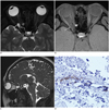
Orbit MRI was performed using 3.0T system (Magnetom Verio; Siemens Medical Solutions, Erlangen, Germany). T2-weighted axial MR images with heavily T2-weighted 3D turbo spin-echo sequence revealed a hyperintense lesion with an internal hypointense linear structure in the right orbital apex (Fig. 1A). No enhancement was noted on gadolinium enhanced T1 weighted axial image (Fig. 1B). The internal structure revealed right oculomotor nerve within the cystic lesion, which continued into oculomotor cistern on oblique sagittal images with heavily T2-weighted 3D turbo spin-echo sequence (Fig. 1C).
The patient underwent right sub-frontal craniotomy and the cystic lesion on prior MR images was shown after unroofing the optic canal. The cystic wall was fenestrated and high pressure cerebrospinal fluid -like fluid was drained. Microscopically, the cystic wall was lined by normal arachnoid membrane, representing arachnoid cyst (Fig. 1D). After the operation, she felt immediate regression of symptoms on the third nerve palsy.
DISCUSSION
The oculomotor cistern (OMC) is arachnoid-lined dural cuff which contains the oculomotor nerve as it enters the cavernous sinus roof. After the oculomotor nerve enters the cavernous sinus, the OMC is gradually tapered and eventually terminates below the tip of the anterior clinoid process (1, 2). Oculomotor nerve penetrates the orbital apex through the cavernous sinus and the superior orbital fissure.
Various pathologic conditions such as schwannoma, cavernous hemangioma, lymphoma, lymphangioma, metastasis, dermoid and epidermoid rarely develop in the OMC. Tanriover et al. (3) reported a case of the schwannoma in the OMC, causing the oculomotor nerve palsy. Itshayek et al. (4) reported another case of cavernous hemangioma of the oculomotor nerve, manifesting facial neuralgia. Diseases involving OMC usually manifest oculomotor nerve palsy due to their anatomic location as in reports previously mentioned.
In general, OMC do not extend into orbital apex because the OMC terminates before reaching the superior orbital fissure. Conditionally, the OMC is able to extend to the orbital apex when the orbital apex syndrome occurs. Orbital apex syndrome is a disease from the orbital apex causing dysfunctions in the optic nerve (II), oculomotor nerve (III), trochlear nerve (IV), abducens nerve (VI), and ophthalmic branch of the trigeminal nerve (V1) (5). Meningocele is one of pathologies involved in this area and have been reported previously (6-9) which were usually meningoceles from optic nerve sheath.
Here, we reported a case of arachnoid cyst arising from oculomotor cistern. Oculomotor cistern is covered by the arachnoid membrane within the dural cuff. Thus, the expansion of oculomotor cistern is able to manifest as dural ectasia or dilation of arachnoid space. In our case, the only arachnoid membrane lined OMC, seemed to be protruding into orbital apex while the dural cuff was terminated at intracranium proximal to orbital apex. It is also observed in type II extradural meningeal cyst with neural tissue at spine (10). To the best of our knowledge, there were no reports about arachnoid cyst of oculomotor cistern at orbital apex.
Arachnoid cysts are fluid-filled cavities within the arachnoid membrane. The symptoms of arachnoid cysts can vary depending on the location and the pressure on surrounding neural structures. So, the oculomotor nerve can be compressed by the arachnoid cyst along any location in its course. Ashker et al. (11) reported a case of an arachnoid cyst compressing the oculomotor nerve in the right interpeduncular cistern. Also, Cheng et al. (12) reported a case of the arachnoid cyst in cavernous sinus, causing deterioration of visual acuity. In most cases, it is sufficient that simple fenestration decompresses arachnoid cyst with decreasing pressure effects.
When a disease occurs in the OMC, precise preoperative evaluation must be performed to prevent injury of the oculomotor nerve. MR with heavily T2-weighted 3D turbo spin-echo sequence can be useful to differentiate various pathologies in the OMC. And, if there is a cystic lesion with signal null on FLAIR image, arachnoid cyst must be included as differential diagnosis.
 XML Download