

 PDF
PDF ePub
ePub Citation
 Print
Print


INTRODUCTION
Desmoid type fibromatosis is a benign fibroblastic tumor, which is previously known as extraabdominal desmoid, aggressive fibromatosis, and musculoaponeurotic fibromatosis (1). Although it is a benign tumor, it tends to be locally aggressive, and infiltrates into the surrounding structures, but does not metastasize (2, 3). It arises from the connective tissue of the muscle, and overlying fascia and aponeurosis. The most common extraabdominal sites include the shoulder girdle, followed by the chest wall and back (1, 2). Involvement of the bone by the tumor is unusual (3-7). Spinal involvements with erosion of the vertebral bodies by paraspinal tumors have been reported (8, 9).
However, to our knowledge, there have not been any reported cases of desmoid type fibromatosis originating from the facet joint of the spine. We report computed tomography (CT) and magnetic resonance (MR) findings of a desmoid type fibromatosis arising from the left facet joint of the lumbar spine in a 31-year-old man treated with partial facetectomy.
CASE REPORT
A 31-year-old man was presented with back pain and mild numbness from the left buttock to the ankle, which developed after the previous motor vehicle accident, 1 year and 5 months ago. Physical examination did not reveal any abnormality in motor or neurological examination. The symptoms were partially improved with epidural and left L5 nerve root block.
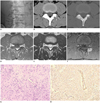
Conventional radiographs of the lumbar spine demonstrated a bony erosive lesion with a sclerotic rim at the left L3-4 facet joint (Fig. 1A). CT revealed an isodense soft tissue mass to the muscle at the posterior portion of the left L3-4 facet joint, which eroded both L3 inferior and L4 superior articular processes, but the articular surfaces of the both eroded facets were well preserved (Fig. 1B, C). MR images demonstrated about 1.6 × 1.0 × 1.0 cm sized soft tissue tumor on the facet joint, which infiltrated beyond the posterior joint capsule. The tumor was isointense to the muscle on T1-weighted image (repetition time [TR]/echo time [TE] 700/10.8 msec) (Fig. 1D), and intermediate to low signal intensity of the tumor between the muscle and cerebrospinal fluid (CSF) on T2-weighted image (TR/TE 4000/97 msec) (Fig. 1E). The lesion was well enhanced by the gadolinium without evidence of the internal necrosis (Fig. 1F). Tc99m-MDP bone scan did not demonstrate an abnormal radiotracer uptake.
The patient underwent bone biopsy, and pathologically, the tumor was diagnosed as a desmoid tumor. After 2 months, the tumor was completely removed with partial facetectomy.
Microscopically, the tumor was composed of cellular proliferation of the elongated, slender, spindle-shaped cells arranged in interlacing bundles of fibroblasts with abundant collagenous stroma (Fig. 1G). The cellularity varied from area to area with scanty mitotic activity, and the cells infiltrated into the adjacent bone. Further, the tumor cells were found in the facet joint capsule with extracapsular infiltration. Immunohistochemical stain showed positive reactions to the smooth muscle actin (Fig. 1H), focal positive reactions to S100 protein, and negative reaction to CD34, vimentin and desmin. The tumor was diagnosed as the desmoid tumor.
After surgical treatment, the patient was relieved from pain in the lower back and buttock.
DISCUSSION
Desmoid type fibromatosis, which is also known as extraabdominal desmoid tumor, musculoaponeurotic fibromatosis and aggressive fibromatosis, is a benign fibroblastic tumor. It arises from the connective tissues, such as the muscles, fascia, aponeurosis or periosteum (1, 5), and may occur in various locations. The most common extra-abdominal sites include the shoulder or upper arm, followed by the chest wall and back, thigh, as well as the head and neck (1-3, 10). Pericapsular location of the tumor was rarely reported in the knee joint (11). In our case, the tumor developed in the facet joint of the lumbar spine.
The incidence of desmoid type fibromatosis is 0.03% of all neoplasms and 3.5% of all fibrous tissue tumors (2, 6). It may occur at any age, but usually in young adults with peak prevalence between the ages of 25 and 35 years (2, 5, 10). In adults, the tumor has a predilection in premenopausal women, but in children, the incidence was equal in both genders. It is manifested as a firm and slowly growing mass. Pain is not typical, unless there is pressure on the surrounding structures. When the tumor occurs in the paraspinal location, scoliosis can be developed with intraspinal extension (8, 9).
Macroscopically, desmoid type fibromatosis is a dense, coarse trabeculated, fibrous tumor with a whorled appearance, and usually infiltrates into fascicles of the skeletal muscles (2, 5, 6). Microscopically, the lesion consists of interweaving the bundles of spindle-shaped fibroblasts in various amounts of collagenous matrix, and the cells lack hyperchromatosia or atypia. Areas of myxoid degeneration and hemorrhage may be seen. The lack of pleomorphism and the low mitotic rate distinguish desmoid from fibrosarcoma. Immunohistochemically, the spindle cells stain with vimentin and smooth muscle actin (1). Our case revealed a positive stain for the smooth muscle actin, but negative for vimentin.
The primary treatment of the desmoid type fibromatosis is a wide resection with negative resection margin (2, 6, 10). The overall recurrence with surgery alone is about 40%. When wide resection cannot be achieved due to infiltrative feature, adjuvant radiotherapy is necessary.
The radiograph of desmoid type fibromatosis is usually normal, or may show a nonspecific soft-tissue mass. Calcification is uncommon. Osseous involvement may be seen in 6-37% of the cases, which manifests as pressure erosion or periosteal frond-like irregularity, and sometimes as cortical destruction and cancellous involvement (4, 5, 10).
CT of desmoid type fibromatosis reveals a soft-tissue mass of the variable attenuation density. The lesions usually show isoattenuation to that of the skeletal muscle, but may be higher or lower with variable amount of collagen matrix or myxoid component. In addition, CT after intravenous contrast enhancement also demonstrates the enhancement of variable degrees. However, there is no relationship between the vascularity of tumor and its histologic features (12). The tumor margin is often indistinct owing to the infiltrative growth pattern.
The MR signal intensities of the desmoid type fibromatosis are variable along with the extent of collagen and the degree of cellularity. Usually, T1-weighted images demonstrate homogeneously similar to slightly hyperintense signal intensity in relation to that of the muscle. On non-fat-suppressed T2-weighted image, the lesions show variable heterogeneous signal intensities. Most lesions show heterogeneous intermediate signal intensity, lower than that of the muscle and higher than that of fat, but less frequently, predominantly low or high signal intensity of the lesions are also found (7, 10, 13-16). The low signal areas on T2-weighted image are related to relatively decreased cellularity and abundant collagen; whereas, the hyperintense areas are correlated with an increased cellularity in the abundant collagen matrix. Prominent low-signal-intensity bands without contrast enhancement are often seen with all pulse sequences and are thought to be the denser areas of the collagen. After injection of gadolinium contrast, the lesions show moderate to strong enhancement, particularly, more in the cellular regions, but less in the collagenous portion. The desmoid type fibromatosis has variable signal intensities on T2-weighted images according to the histologic evolution (10, 16). The initial lesions are more cellular with large extracellular spaces and less areas of hyalinized collagen, which demonstrate increased signal intensity. As the amount of collagen deposition is increased in the central and peripheral areas, the signal intensities of the lesions become variable. Finally, as the fibrous compositions are predominant with a decrease in cellularity and extracellular spaces, the tumors overall appear as low signal intensity. Our case revealed intermediate to slightly high signal intensity on T2WI, which was correlated with typical cellularity with collagenous stroma.
Differential diagnoses of our case, arising from the facet joint include collagenous fibroma, nodular synovitis, and pigmented villonodular synovitis (PVNS). Collagenous fibroma (desmoplastic fibroma) is a benign fibrous soft tissue tumor, which is composed of spindle shaped fibroblasts embedded in a dense fibrous stroma. The tumor mostly involves subcutaneous fat or deep soft tissue. Vertebral involvement with bony erosion (17) and intraarticular location in the hip joint (18) have been reported. The nodular fasciitis is a benign myofibroblastic proliferating tumor-like lesion with a predilection for the subcutaneous tissue, fascia and within the muscle. Intraarticular location of the nodular fasciitis has been also reported (19, 20). The CT and MR imaging findings of the nodular fasciitis are nonspecific, according to the amount of cellularity and fibrous component. PVNS is a benign, local aggressive lesion, which is characterized by villous or nodular proliferation of the synovium in the joints, bursae or tendon sheaths. PVNS usually involves the knee and hip joint. Involvement of the facet joint by PVNS with bony erosion of the adjacent lamina is rarely reported (21, 22). These tumors cannot be differentiated by imaging findings alone.
In conclusion, desmoid type fibromatosis can involve the facet joint, and should be considered in the differential diagnosis of the fibrous soft tissue tumor in the facet joint of the spine.
 XML Download