

 PDF
PDF ePub
ePub Citation
 Print
Print


INTRODUCTION
In general, more common anomalies of renal vein include the left circumaortic renal vein, left retroaortic vein and multiple renal veins. Several former studies demonstrated the prevalence and clinical implications of these renal venous anomalies focusing on the left renal vein (1-3). Right renal variations rarely occur due to relatively simple embryological development, as compared to its left counterpart. To the best of our knowledge, multi-detector CT (MDCT) findings concerning the right circumaortic renal vein have never been reported. We present an extremely rare case with the circumaortic route of the right renal vein and right ectopic kidney. Precise awareness of a rare renal venous variation is important for accurate diagnosis in the staging of urologic cancers and helps prevent unforeseen complications during operations or interventional procedures.
CASE REPORT
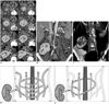
A 42-year-old man underwent abdominopelvic MDCT due to the evaluation for traumatic splenic rupture. CT examination was performed using a 128-detector-row CT scanner (definition AS+, Siemens Medical Solutions, Forchheim, Germany). Helical scan data was acquired using 32 × 1.2 mm collimation, a rotation speed of 0.5 sec, pitch of 0.6, and 120 kVp. Attenuation-based tube current modulation was used with a reference tube current-time product set at 210 mAs per rotation. Volume rendering images were obtained by using Aquarius iNtuition viewer (Terarecon, SanMateo, CA, USA). A right ectopic kidney was found incidentally. The left kidney was in an orthotropic position with normally coursing the renal vein. The right ectopic kidney was located between the level of the 3rd lumbar vertebral body superiorly and 5th lumbar vertebral body inferiorly. The right ectopic kidney was incompletely rotated while the hilum and renal pelvis were facing anterior to the parenchyma. The anterior portion of the right renal vein which was emerging from the anterior part of the ectopic kidney crossed to the left side of the aorta with a ventral course to the inferior vena cava (IVC) and aorta (Fig. 1A, C). Then, it turned around and coursed dorsally to the aorta and finally drained into the IVC (Fig. 1A, C). The right renal artery arose from the abdominal aorta as the upper polar artery (Fig. 1A). The traumatic splenic lacerations and hemoperitoneum are also noted (Fig. 1B). The patient showed American Association for the Surgery of Trauma splenic injury grade 2 and stable vital signs, so conservative treatment was performed.
DISCUSSION
The development of the renal vein is closely related with the embryogenesis of the IVC (1, 2). The IVC is formed from three parallel veins that subsequently appear and regress (Fig. 1D). In order of appearance, they are the posterior cardinal veins, the subcardinal veins and the supracardinal veins. First, the posterior cardinal veins develop and receive blood returning from the body below the heart. Next, the subcardinal veins appear by the fifth week. The subcardinal veins lie in a plane ventral to the aorta. Lastly, the supracardinal veins develop, and it lies in a plane dorsal to the aorta. The subcardinal veins and the supracardinal veins are interconnected by a network of veins, forming a venous collar around the aorta. The renal veins are formed by the anastomoses of the supracardinal and subcardinal veins. Two renal veins form as ventral and dorsal; the dorsal vein usually degenerates, the ventral vein forms the renal vein.
Various anomalies of the renal vein can develop during the developmental process (3). The most common anomaly is the circumaortic left renal vein, which results from persistence of the dorsal vein and intersupracardinal anastomosis. The prevalence of circumaortic left renal vein is up to 8.7%. Another common anomaly is a retroaortic left renal vein, which has a prevalence of 2.7%. As with a circumaortic left renal vein, a retroaortic left renal vein results from persistence of the dorsal arch of the renal collar. However, in this variation, the intersubcardinal anastomosis and ventral vein regress so that a single renal vein passes posterior to the aorta. In contrast to the left circumaortic and retroaortic vein, abnormal course of the right renal vein is extremely rare (4). The development of the left renal vein is more complex than its right counterpart. In our case, the circumaortic route of the right renal vein was formed by the persistence of the intersubcardinal anastomosis, left suprasubcardinal anastomosis and the intersupracardinal anastomosis (Fig. 1E). The intersubcardinal anastomosis formed the ventral arch of the right circumaortic renal vein, while the intersupracardinal anastomosis formed the dorsal arch. The left suprasubcardinal anastomosis connected between the ventral and dorsal arch at the left paraaortic area.
Anomalies of renal vessels are particularly frequent in kidneys that do not ascend or rotate normally. Besides the circumaortic and retroaortic renal veins, there are supernumerary veins, double renal vessels and accessory renal vessels related to ectopic kidneys that have been reported.
Renal venous anomalies have several important clinical implications in retroperitoneal surgeries (5). Since anomalous venous structures tend to be dilated and tortuous, causing injury more easily, lack of preoperative recognition of renal vein anomalies can have potentially disastrous consequences (6). Intraoperative trauma of an anomalous vein may cause life-threatening hemorrhage. The relationship of the ureter to the vessel in the retroperitoneum may also be confusing. Control of the proximal aorta may be difficult to achieve in patients with circumaortic collars. During removal of donor kidneys or urological procedures, veins require close attention because venous variations may lead to severe vascular complications. It is also important for radiologists to interpret angiograms or staging for lymph node evaluation (6). The right circumaortic renal vein is seen in the left retroperitoneal space, so surgeons may misconceive it as the left renal vein.
 XML Download