

 PDF
PDF ePub
ePub Citation
 Print
Print


INTRODUCTION
Hemangioma may occur in any region of the body but it typically presents as cutaneous lesions of the head, neck and trunk. Visceral hemangiomas have been described in various organs, and they are common in the liver. Pancreatic hemangioma is very rare, especially in adults. Only a few cases of adult pancreatic hemangiomas have been reported in the English literature (1-6). They typically appear on computed tomography (CT) as a cystic lesion with contrast enhancement in the arterial phase. Herein, we report a case of adult pancreatic hemangioma. The mass showed a multilocular cyst with fluid-fluid levels and no obvious enhancement on CT and magnetic resonance imaging (MRI) whereas previously reported cases showed intense enhancement on CT after injection of contrast material, and high signal intensity on T2WI on MR (1-6).
CASE REPORT
In a 23-year-old woman with no symptoms, a 5.4 × 5.0 × 3.1 cm mass with internal heterogeneous hyperechogenicity in the pancreas head was found during an annual ultrasound health examination. Blood test findings were within normal range, including the complete blood count, liver function tests and tumor markers. The physical examination revealed no significant abnormality.
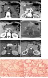
CT was performed on a GE Lightspeed VCT scanner and included a plain scan and dual-phase enhanced scans respectively. CT parameters included 64 × 0.625 mm detector collimation, 120 kVp, 180 mAs, 1.0 pitch, 5 mm slice thickness. An enhanced scan using Iohexol (GE Healthcare, Shanghai, China) contrast agent during the arterial phase and portal venous phases (at 32 and 70 seconds, respectively, dose 1.5 mL/kg, injection flow rate 3.0 mL/s) were also performed. A plain CT scan of the abdomen showed a heterogeneous, well-defined mass at the pancreatic head. A contrast-enhanced CT scan revealed a heterogeneous, nonenhancing, multilocular cyst with fluid-fluid levels at the pancreatic head. The lesion grew outward and extended into the lesser sac (Fig. 1A-C).
MR imaging was performed with a SIEMENS 1.5-T superconducting system with a body coil. Sequences included T2-weighted fast spin echo with fat suppression (repetition time/echo time, 3250/110 msec; section thickness, 5 mm; gap, 1 mm) and T1-weighted breath-hold gradient echo (repetition time/echo time, 160/4.9 msec; section thickness, 5 mm; gap, 1 mm). A T1-weighted gradient-echo with fat suppression was performed during the hepatic arterial and portal venous phases (at 20 and 50 seconds, respectively) after manual administration of 0.1 mmol/kg Gadodiamide (GE Healthcare, Dublin, Ireland). The MR findings were similar to the CT findings. On MR, the superior fluid layer showed hyper signal intensity (SI) on T1WI and T2WI. The inferior fluid layer was slightly hyper SI to the pancreas on T1WI and hypo SI relative to the superior fluid layer on T2WI (Fig. 1D-F).
Subtotal pancreatectomy was done. At laparotomy, a soft, blood-red tumor was found arising from the head of the pancreas. There was no invasion of the surrounding vascular structures and the duodenum. The mass was resected from the pancreas. Gross pathologic examination revealed a reddish tumor composed of multiloculated cysts containing intracystic hemorrhaged blood. Microscopically, the tumor showed multiple vascular cysts of variable size lined by a single layer of uniformly flattened cells. The vascular cysts were separated by fibrous tissue (Fig. 1G, H). Histologic examination was consistent with pancreatic hemangioma. The patient was healthy without any sign of recurrence oneyear after surgery.
DISCUSSION
Typically, pancreatic hemangiomas of previously reported adult cases were reported as well-circumscribed with homogeneous lower density and showing intense enhancement after injection of contrast material on CT (1-6). According to the only report of its MR findings (1), pancreatic hemangiomas were reported as lesions with iso SI on T1WI and hyper SI on T2WI, similar to cavernous hemangiomas. In our case, however, the imaging findings were completely different to those of previous reports, which described multilocular cysts with fluid-fluid levels and no obvious enhancement.
Mulliken et al. (7) divided the natural history of hemangiomas into three phases based on physical findings and cellular features: proliferation, involution and involuted. Histologically, multilaminated basement membrane formation beneath the endothelium in the proliferative phase turns into a single layer of uniform and flattened cells with fibrosis and fat deposition in the involuted phase. The microscopic findings of our case were consistent with those of the involuted phase. Moreover, MRI is the preferred cross-sectional imaging technique for differentiating the different phases of a hemangioma a (7). In the proliferative phase, hemangiomas demonstrate intermediate SI on T1WI and moderate hyper SI on T2WI. Especially, flow voids in and around the tumor due to the fast flow and vascular shunting can be seen. In the involuted phase, hemangioma shows decreased flow voids within the tumor. The flow voids in and around the lesion of our case were not displayed on MRI. Therefore, the multilocular cyst in our case may have been related to the involuted phase. The lesion in our case rapidly expanded in the proliferative phase and subsequently a multilocular cyst formed in the involuted phase. Meanwhile, the ratio of the cystic to solid component can influence the degree of tumor vascularity (3). The lesion of our case principally manifested as a multilocular cyst. So, slow blood flow due to the multilocular cyst resulted in no obvious enhancement after injection of the contrast material in the CT and MRI in our case.
The fluid-fluid level demonstrates that the lesion contains different fluids of different densities so that a sedimentation effect can occur, and can be demonstrated on CT and MR imaging, provided that the plane of the scan is perpendicular to the horizontal (8). Fluid-fluid levels should not be considered diagnostic, as this finding may be present in various other histologic tumors as well. Fluid-fluid levels occurring in hemangiomas of soft tissue and liver have been described in the literature (8, 9). However, to our knowledge, we are the first to describe the CT and MRI findings of fluid-fluid levels in a pancreatic hemangioma. It is well known that fluid-fluid levels in various histologic tumors are attributable to cystic change or hemorrhage. In our case, the lesion was composed of multiloculated cysts containing intracystic hemorrhage. Therefore, we thought that the stagnant or slowly flowing blood resulted in the sedimentation effect, with unclotted serous blood constituting the superior fluid layer and sedimentary red blood cells constituting the inferior fluid layer. This hypothesis was supported by CT and MR imaging in our case. Unclotted serous blood shows characteristics of a fluid on the CT and MRI. Red blood cells of the inferior fluid layer demonstrated higher attenuation on CT, iso SI to muscle on T1WI and hypo SI relative to the superior fluid layer on T2WI.
The radiological appearances of a multilocular cyst and the fluid-fluid levels in our case should be differentiated from those of other multilocular cystic tumors of the pancreas, including pseudocysts, serous cystadenomas, mucinous cystadenomas and side-branch type intraductal papillary mucinous neoplasms (IPMN). A fluid-fluid level is a very rare appearance in the above mentioned tumors; however, hemorrhage has occasionally been reported when the tumor bleeds into itself (10). A possible mechanism of hemorrhage in pancreatic pseudocysts is erosion of the pseudocyst into a vessel due to persistent compression and ischemia, and the elastolytic actions of enzymes. Most patients with pseudocysts and accompanying hemorrhage have a ruptured intracystic pseudoaneurysm, which is readily diagnosed as a rapidly enhancing intracystic lesion with attenuations similar to those of the aorta on CT (11). The CT appearances of serous cystadenoma and mucinous cystadenoma are similar. A serous cystadenoma appears as a multicystic or lobulated cystic lesion with septation, while a mucinous cystadenoma has a smooth shape, with or without septations. Calcification in serous cystadenomas is typically central within the fibrous stroma, whereas a mucinous cystadenoma may have peripheral eggshell calcification. Side-branch type IPMN appears as a pleomorphic cystic mass. An imaging diagnosis of side-branch type IPMN depends on identifying the communication between the lesion and the main pancreatic duct (12).
Conclusions
Pancreatic hemangiomas in adults are very rare. Although multilocular cyst, fluid-fluid levels and no obvious enhancement are three findings in our case compared with the previously reported cases, knowledge of the presence of these findings might help in determining the nature of the pancreatic hemangioma and may help in confirming the diagnosis.
 XML Download