

 PDF
PDF ePub
ePub Citation
 Print
Print


INTRODUCTION
Mucosa-associated lymphoid tissue (MALT) lymphomas are the extranodal subset of marginal zone B-cell lymphomas and represent less than 8% of all of types of lymphomas (1). MALT lymphomas were initially described in 1983 by Isaacson and Wright as a distinctive type of B-cell lymphoma arising in the gastrointestinal tract (2). Since this original description, numerous publications have documented their occurrence in various organs, including the stomach, small bowel, lung, thyroid glands, and salivary glands (3, 4). However, primary occurrence in the common bile duct (CBD) is extremely rare and, to our knowledge, only four cases have been reported in the English-language literature (3, 5-7). We report an extremely rare case of primary low-grade B-cell lymphoma of the MALT type arising from the CBD which clinically and radiologically mimicked cholangiocarcinoma on the preoperative evaluation.
CASE REPORT
A 79-year-old male presented with a four-week history of vague, generalized abdominal discomfort and progressive jaundice. He complained of pruritus and dark urine, although he showed no weight loss, febrile sensation or chills. He had no history of biliary stone disease or prior abdominal surgery. Physical examination revealed jaundice with icteric sclera. Laboratory studies showed cholestasis with a total bilirubin of 2.2 mg/dL (normal, < 1.0 mg/dL), direct bilirubin of 1.5 mg/dL (normal, < 0.3 mg/dL), aspirate amino transferase of 141 IU/L (normal, < 40 IU/L), alanine transferase of 190 IU/L (normal, < 56 IU/L), and alkaline phosphatase of 284 IU/L (normal, < 147 IU/L). Serum tumor markers, including CEA and CA 19-9, were not elevated.
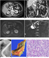
Computed tomography (CT) examination was performed on a 16-multidetector CT system (LightSpeed 16; GE Healthcare, Milwaukee, WI, USA) with triphasic dynamic imaging. The arterial-, portal-, and delayed-phase images were obtained using a 25-s, 72-s, and 180-s delay, respectively, after intravenous injection of 2.5 mL/kg of iopromide (Ultravist 370; Bayer Schering Pharma, Berlin, Germany) at a rate of 3 mL/s. The CT parameters were beam collimation of 16 × 1.25 mm, beam pitch of 0.938, 120 kV, 200 mAs, and slice thickness of 3 mm. CT revealed diffuse, irregular wall thickening of the CBD, including the intrapancreatic portion of the CBD, with minimal dilatation of the common hepatic duct (CHD) and the intrahepatic duct (IHD) (Fig. 1A, B). The thickened ductal wall showed relatively homogeneous enhancement with isoattenuation compared to the liver parenchyma on the arterial, portal, and delayed phases. A small lymph node with a short-axis diameter of 8 mm was observed within the hepatoduodenal ligament. Magnetic resonance imaging (MRI) with MR cholangiography using a 1.5 T scanner (Magnetom Avanto; Medical Solutions, Erlangen, Germany) depicted ductal wall thickening with slight hypointensity relative to the liver parenchyma on T1-weighted images (TR/TE, 164/4.5; flip angle, 70°; FOV, 350 × 250; slice thickness, 6 mm) and slight hyperintensity on T2-weighted images (TR/TE, 1900/88; echo train length, 13; flip angle, 150°; FOV, 350 × 250; slice thickness, 6 mm). Diffusion-weighted images with a b-value of 800 showed heterogeneous hyperintensity in the CBD (Fig. 1C). Although the distal CBD lumen was relatively maintained, the entire CBD exhibited irregular luminal narrowing on MR cholangiography using single-shot rapid acquisition with relaxation enhancement sequence (TR/TE, infinite/1000; echo train length, 256; FOV, 250 × 250; slice thickness, 40 mm) (Fig. 1D). The CHD and IHD showed mild dilatation, which was in agreement with the CT findings.
In order to relieve the patient's obstructive jaundice, we performed endoscopic retrograde cholangiopancreatography (ERCP) with plastic biliary drainage catheter placement. ERCP revealed diffuse, irregular luminal narrowing of the entire CBD, including the intrapancreatic portion, with diffuse, mild dilatation of the upstream biliary tree (Fig. 1E). ERCP also showed an out-pouching, diverticulum-like lesion in the proximal CBD.
A preoperative diagnosis of cholangiocarcinoma involving the CBD was made, and the patient underwent pyloruspreserving pancreaticoduodenectomy. Grossly, the CBD was found to be diffusely thickened (Fig. 1F). No significantly enlarged regional lymph nodes were found, although routine lymph node dissection was performed. Histologic examination under light microscopy revealed dense and diffuse infiltration of atypical lymphoid cells and the formation of lymphoid follicles within the CBD walls (Fig. 1G). On immunohistochemical staining, the tumor exhibited CD20 positivity and CD3 negativity. Ki-67 was almost negative with less than 1% positivity. Lambda, kappa, IgG, and IgG4 were all noncontributory. A final pathology diagnosis of low-grade B-cell lymphoma of the MALT type arising from the CBD was made. Of the 15 dissected lymph nodes, no foci of lymphoma involvement were discovered.
DISCUSSION
Mucosa-associated lymphoid cells are normally found in the epithelium of the different mucosae of the body. They serve primarily an immunologic function, and IL-2 is known to be an important mediator of the immunologic response (8). However, in the presence of chronic inflammation, prolonged lymphoid reactive proliferation occurs and leads to irreversible genetic rearrangements, making the cells unresponsive to IL-2 regulation. Without control of IL-2 regulation, MALT lymphoma develops (9).
Primary lymphoma involvement of the bile duct is rare and is usually a secondary involvement of disseminated abdominal lymphomatous disease (6). Since Nguyen reported the first case of primary CBD lymphoma in 1982, only 23 cases of primary CBD lymphoma have been reported in the medical literature (10, 11). Although various lymphoma subtypes have been reported in the literature, the most common subtype is large-cell lymphoma and the most common immunophenotype is B-cell lineage (12, 13). On the other hand, MALT lymphoma of the CBD is extremely rare, and our extensive review of the medical literature disclosed only four previously reported cases (3, 5-7). All four of these cases were elderly patients (age, 62-76 years) with an equal gender distribution.
In the four reported cases of MALT lymphoma of the CBD, three cases demonstrated diffuse, concentric CBD wall thickening without a definite mass on CT, while the other cases appeared as a focal, lumen-obstructing mass within the CBD on CT. However, only one case (7) showed the detailed imaging findings of MALT lymphoma of the CBD as thickened CBD wall with homogeneous enhancement on CT, slight hyperintensity on T2-weighted MR images, and hypointensity on T1-weighted MR images. In our patient, pathologic wall thickening showed similar findings on CT and MRI to those of the previous case. However, in the previous case (7), the authors proposed a characteristic feature of primary biliary lymphoma as they found a discrepancy in the degree of luminal narrowing between the cross-sectional examinations, including CT or MRI, and ERCP. Although CT and MRI demonstrated diffuse luminal narrowing with thickening of the extrahepatic bile duct and upstream IHD dilatation, ERCP revealed relatively subtle, symmetric luminal narrowing. They argued that because lymphomas are usually not associated with desmoplastic reactions, the degree of luminal narrowing and mucosal irregularities may be less prominent in lymphomas than in cholangiocarcinomas (14). However, as a large margin for error remains, such disagreement requires further validation. Although such a discrepancy between cross-sectional examinations and ERCP was not exhibited for our patient, the overall degree of CHD and IHD dilatation was not as severe as that normally expected with cholangiocarcinoma. However, as upstream bile duct dilation can be minimal in some cases of cholangiocarcinoma, the differential diagnosis may be even more difficult. Owing to such difficulties, the vast majority of primary CBD lymphoma cases, including our case, are diagnosed only after surgical acquisition of adequate pathologic specimens.
In our case, ERCP revealed a diverticulum-like structure within the proximal CBD. Along with biliary webs, biliary tree diverticula were once considered as well-recognized cholangiographic features of primary sclerosing cholangitis (PSC) and were believed to be specific for this disease (15). However, more recent studies concluded that these features are nonspecific for PSC and can be seen in patients with choledocholithiasis, cholangitis, biliary stricture (benign or malignant), biliary stents with balloon dilatation, and biliary diversion (16). Nevertheless, as this ERCP feature was not seen in the four previously reported cases of MALT lymphoma of the CBD, the significance of such finding should be further evaluated.
A correct early diagnosis of primary lymphoma of the CBD and its differential diagnosis from the much more common cholangiocarcinoma are very difficult. As with cholangiocarcinoma, the most common manifestation of the total 23 reported cases of primary CBD lymphoma is acute onset of obstructive jaundice with or without abdominal pain. However, as primary lymphoma accounts for less than 1% of all biliary obstructions and there is a general lack of familiarity with the diagnosis, primary lymphoma is seldom considered in the differential diagnosis (5, 12). Nonetheless, the dramatic difference in the prognosis and treatment of cholangiocarcinoma and primary lymphoma emphasizes the importance of the differential diagnosis in extrahepatic bile duct tumors with obstructive jaundice. To date, there are no known characteristic radiologic findings specific for the diagnosis of primary lymphoma of the CBD, including MALT lymphoma (3-7, 10, 12, 17, 18). CT scans in previously published reports demonstrated diffuse wall thickening of the CBD with or without the presence of an intraductal mass lesion causing proximal bile duct dilatation. A thickened bile duct wall and an intraductal mass exhibited enhancement upon intravenous contrast administration. In addition, although reports regarding the MR imaging findings are limited, the pathologically thickened bile duct wall shows hyperintensity on T2-weighted images and hypointensity on T1-weighted images. However, the CT and MRI findings described above are also consistent with those of cholangiocarcinoma, thus making a correct preoperative diagnosis challenging.
In conclusion, we report an extremely rare case of primary low-grade B-cell lymphoma of the MALT type arising from the CBD, causing obstructive jaundice at the time of the initial presentation. As the clinical and radiological findings of CBD MALT lymphoma resemble those of cholangiocarcinoma, an accurate radiologic differential diagnosis is extremely difficult.
 XML Download