

 PDF
PDF ePub
ePub Citation
 Print
Print


INTRODUCTION
Amyloidosis whether generalized or localized can affect the breast. By definition, amyloidosis is the extracellular deposition of amorphous congophilic protein within tissues. Many reports have described the image findings of primary breast amyloidosis as a mass on mammography. Breast amyloidosis manifesting only as calcifications have been rarely reported (1-5). Herein, we report a case of localized amyloidosis of the breast presenting suspicious microcalcifications which are not associated with a mass as seen on the mammography.
CASE REPORT
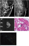
A 72-year-old woman was referred to our hospital for further evaluations of calcifications detected on a screening mammography taken at another hospital. The patient had a 5-year history of diabetes mellitus. No other abnormality was evident from basic laboratory examinations. No breast symptoms were evident such as pains, palpable masses, or nipple discharges. Mammography demonstrated multiple, irregular calcifications in the subareolar area of the left breast; they were regionally distributed, generally smooth-branched, linear, and rod-like, and varied in sizes and shapes (Fig. 1A, B). These calcifications were classified as category "4A": low suspicious findings at final assessment according to the Breast Imaging Reporting and Data System (6). Thus, a biopsy was recommended and the patient underwent surgical excisional biopsy with mammoguided needle localization. Specimen mammography revealed several irregular-shaped calcifications (Fig. 1C). There were no discrete mass evidences on the gross pathology. Histopathology examination revealed dystrophic calcifications in ectatic mammary ducts, amorphous eosinophilic material deposition; the infiltration of periductal lymphocytes and multinucleated giant cells were revealed upon hematoxylin-eosin staining (Fig. 1D). The calcifications were stained with Congo-red and exhibited apple-green birefringence under polarizing microscopy (Fig. 1E) which were consistent with amyloidosis. No malignancies were pathologically identified. Immunohistochemistry confirmed the AL type amyloidosis. Our patient refused further investigations, and there was no clinical evidence suggesting generalized amyloidosis in her physical or laboratory results. There was no interval change in calcifications of the left breast on follow-up mammography for five years. During the follow-up period of five years, no clinical or laboratory evidence of systemic amyloidosis has been demonstrated.
DISCUSSION
Amyloidosis is the deposition of amyloid at connective tissues framework of organs. It is divided into the systemic and localized form, according to the extensions of the disease and it can be classified as a primary or secondary form according to the etiology. Amyloidosis can also be classified as the AA or AL type based on chemical compositions. In clinical practice, the most commonly diagnosed form of amyloidosis is primary idiopathic amyloidosis, the AL type (7, 8). Breast amyloidosis typically appears as diffuse breast involvements in the systemic form of amyloidosis with the primary AL type rather than AA type (8). A less common manifestation is the isolated breast involvement that occurs as a localized form of disease (7, 8). The clinical course tends to be benign, and typically, patients complain for hard, non-painful, and palpable masses in the affected breasts (5). Sometimes, a patient may present abnormalities on the mammograms without any clinical symptoms. Previous studies have reported that common mammographic findings of breast amyloidosis have a variety of solid shapes or multiple masses or nodules, whether including or excluding calcifications (9, 10). Primary breast amyloidosis which consists of only microcalcifications excluding the associated mass is very rare. Only five cases that were presented solely as microcalcifications have been reported (1-5). One case was breast involvements of systemic amyloidosis within a patient with a 15-year history of multiple myeloma (3). The remaining four reports were in localized forms. In the previous reports, the shapes of the microcalcifications varied according to clustered (4, 5), pleomorphic (4, 5), fine linear and branching (1, 2), and smooth branching rod-like (2, 3) shapes. All the previous mammographic findings were assessed either as intermediate or suspicious of malignancy, and pathological confirmations were recommended (1-5). In this case, there were irregularly linear distributed, multiple, smooth, rodlike calcifications as evident by mammography, similar to the two previously reported cases (2, 3). Among these two prior reports, one is of localized form, and the other being breast involvements of systemic amyloidosis (2, 3). This implies that mammographic findings between systemic amyloidosis and localized amyloidosis are not different which is probably due to the same pathophysiology. In breast amyloidosis, the amyloid is histologically evident as depositions at periductal, interstitial, or perivascular spaces with multiple multinucleated giant cells and calcifications. Amyloid fibrils have an affinity for calcium and deposition around mammary ducts and is found in blood vessels (7), thus, this pathophysiology is strongly correlated with the mammography-evident branching or linear distribution of microcalcifications deposited in or around the vasculature or mammary ducts. Few cases of breast amyloidosis were associated with breast cancers such as ductal carcinoma in situ, invasive carcinoma, invasive lobular carcinoma, or tubular carcinoma (8-10). And, some cases were associated with diffuse skin thickening which mimics inflammatory breast cancer (10). In our case, no malignancy was identified on pathology. Although no distinctive findings have been reported for breast amyloidosis, the previously reported cases presenting suspicious masses with or without microcalcifications that mimic malignancy and pathologic diagnosis were needed.
In summary, breast amyloidosis is a rare entity that is usually evident as clinical or radiological palpable masses. Breast amyloidosis which only presented microcalcifications without mass is still rare, and only a few cases have been reported (1-5). Herein, we report a case of breast amyloidosis detected upon mammography which was solely presented as suspicious microcalcifications without mass as diagnosed by surgical excisions.
 XML Download