

 PDF
PDF ePub
ePub Citation
 Print
Print


INTRODUCTION
Advances in transducer design, electronics, computers, and signal processing have improved the quality of ultrasound images to the extent that ultrasonography is now an important imaging modality for the clinical diagnosis of breast cancer (1-4). These developments, along with the establishment of a standardized lexicon for features of solid masses, have improved the diagnostic performance of breast ultrasounds (4).
In most breasts, fat lobules are embedded within the fibroglandular tissue. Therefore, misclassification of isoechoic true lesions as fat or fat as isoechoic nodules can frequently occur in breast ultrasounds (5). In particular, band-like interposing fat is one of the pseudolesions that are misinterpreted as a malignant lesion according to standardized Breast Imaging Reporting and Data System (BI-RADS) lexicon and category classification, often resulting in an unnecessary biopsy. Band-like interposing fat is defined as fat lobules with a radial or not parallel orientation that cross between the fibroglandular tissues. Large vessels, such as the lateral thoracic artery or its branches, could be located in or adjacent to the band-like interposing fat. When this anatomical relationship is not recognized, the lesion could be diagnosed as a hypervascular mass. Pathology results often confirm that biopsies of such samples represent normal tissue and fat. Large vessels are occasionally included in histopathological examinations.
The aim of this study was to examine the clinical characteristics and ultrasonographic findings of band-like interposing fat, which is an ultrasonographic pseudolesion that occurs in the breast, as well as to analyze how it is interpreted by breast radiologists and to suggest additional approaches for its diagnosis.
MATERIALS AND METHODS
Our institutional review board approved this retrospective study and waived informed patient consent. We performed a retrospective review of the ultrasonographic and histopathological findings in patients with breast lesions of BI-RADS category of 4 or higher which had been confirmed as benign between June 2008 and June 2010. At our institution, we performed ultrasonography-guided core biopsies in 3125 patients between June 2008 and June 2010. Of those patients, 1391 patients had a lesion of BI-RADS category greater than 4. Benign breast lesions were confirmed in 804 of the 1391 cases. A pathologist reviewed the specimens that were reported as normal fat with normal tissue, normal fat with a large vessel, simply normal fat, and nonspecific fibrocystic changes with a sonographically suspicious band-like interposing fat lesion. After review by the breast pathologist, we included 26 lesions in this study. According to the protocol, 22 category 4a lesions were confirmed based on the ultrasonography-guided core needle biopsy and were followed-up by regular imaging. Additionally, 4 lesions that were greater than category 4b were confirmed upon repeated ultrasonography-guided core needle biopsy (n = 2), vacuum-assisted biopsy (n = 1) or surgery (n = 1) and were followed-up by regular imaging. All lesions were followed up for 24-48 months. We performed an analysis of the clinical characterization of these lesions, including symptoms, location, and size.
HDI 5000 and iU22 (Ultrasound system, Philips ultrasound, Bothell, WA, USA) ultrasound machines were used in this study. Two breast radiologists with 5 and 7 years of experience examined the images and selected the best images for analysis.
Five well-trained radiologists with six to twelve years of experience were asked to analyze the cases. The radiologists who selected the ultrasound images were not included as reviewers. They described and assessed the images according to BI-RADS lexicon and category classification. The reviewers were blinded to the clinical information and were asked to analyze the axial and sagittal ultrasound and Doppler images based on BI-RADS classification.
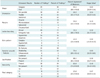
We evaluated the ultrasound findings in the 26 cases. The five radiologists selected the most appropriate characteristics of each lesion as described by the BI-RADS (shape, orientation, margins, interface, echo pattern, posterior acoustic feature, calcification, vascularity, and final category). First, we assessed the consensus (an agreement of 3 or more of the 5 reviewers) results of the 26 cases and the most frequent findings. Second, to calculate the interobserver reliability and to determine the degree of consistency for each lexicon between observers, the percent agreement and the kappa statistical analysis function of SAS version 8.0 (MAGREE SAS Macro program, SAS Institute Inc., Cary, NC, USA) were used. Kappa values for multiple observers and multiple lexicons were calculated based on the methods proposed by Fleiss. According to the k value, the degree of agreement is slight (k ≤ 0.20), fair (0.21 ≤ k ≤ 0.40), moderate (0.41 ≤ k ≤ 0.60), substantial (0.61 ≤ k ≤ 0.80), or near perfect (0.81 ≤ k) (6). Third, we evaluated the differences in the interpretation of the ultrasonographic findings relative to the experience of the observer. Finally, we correlated the ultrasonographic findings with the findings from other imaging modalities, such as mammography or breast MRI. Mammography was performed on 21 of the 26 patients. MRI was performed in one patient.
RESULTS
The mean age of the 26 patients was 43.7 ± 9.9 years (range: 24-58 years). All of the 26 patients were asymptomatic, and the lesions were identified during either the screening or follow-up evaluation for other likely benign lesions.
There were right breast lesions in 7 cases (26.9%) and left breast lesions in 19 cases (73.1%). All seven lesions in the right breast were located between the 9 and 12 o'clock position of the breast (the upper outer quadrant). Of the 19 left breast lesions, 17 were located in the upper outer quadrant, and the remaining two were located in the upper inner quadrant. Thus, 24 (92.3%) of the 26 lesions were located in the upper outer quadrant. The mean distance from the nipple of these lesions was 2.4 ± 0.7 cm (1.1-4.5). Though the distance of the lesions from the nipple varied, they did not occur near the nipple or the axillary tail. The mean depth of the lesions from the skin was 1.3 ± 0.3 cm (0.8-2.1), occurring neither as superficial nor deep but instead confined to the mid parenchyma area. The maximal length of these lesions was small, with a mean of 0.8 ± 0.4 cm (0.3-1.8).
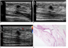
The consensus ultrasound findings and the most frequent findings in each of the nine lexicons were determined for the 26 cases (Table 1). We used the percentage agreement to examine the interobserver reliability and the kappa statistic in order to determine the degree of consistency for each lexicon between observers. The results are presented in Table 1. We found that the level of experience of the observer was positively correlated with the increased tendency to identify lesions as benign or normal rather than malignant. Among the five radiologists, one with 12 years of clinical experience interpreted 17 cases as benign (65.4%). One radiologist with 10 years of clinical experience interpreted 15 cases as benign (57.7%). In particular, 11 of 15 of these cases corresponded to normal findings in category 1. One radiologist with 8 years of clinical experience interpreted 10 cases as benign (38.5%). The last two radiologists, each with 6 years of clinical experience, interpreted all the lesions as malignant (0%). However, there were five cases that were interpreted as malignant by all five readers (Fig. 1), five cases that were interpreted as malignant by four readers, 11 cases that were interpreted as malignant by three readers, and five cases that were interpreted as malignant by two readers.
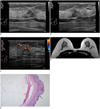
There were no suspicious findings detected by mammography. In the MRI scans, the interposing fat was observed without any suspicious lesions (Fig. 2).
DISCUSSION
Although breast cancer is the most common cancer in women, mammography is the only screening tool that has been shown to reduce mortality (7, 8). Mammography for breast cancer screening in younger women has been controversial because breast tissue in these patients has high parenchyma density that can obscure the lesions and decrease the sensitivity of mammography (9, 10). Several reports have concluded that breast ultrasound is useful in dense breast tissue (2, 11, 12). Although breast ultrasound is used for breast cancer screening, the lower specificity of the ultrasound compared to the mammography is a limitation (1-3). The specificity of the breast ultrasound will be markedly improved if it can reliably differentiate between true lesions and pseudolesions during screening.
Common pitfalls of breast ultrasound include the failure to discriminate the sonographic appearance of normal breast anatomy, such as the rib, the nipple, fat lobules, lactiferous ducts, or acoustic shadowing of Cooper's ligament. To prevent unnecessary biopsies, it is important to recognize the potential artifacts and pitfalls (13).
At our institution, full-field digital mammography is a routine screening tool. In women with dense breast tissue, combined mammography and ultrasonography is performed for screening. In such cases, some types of lesions can only be detected by an ultrasound, including likely benign lesions, small breast cancers, and pseudolesions, such as band-like interposing fat. In the current study, we found 26 confirmed cases of band-like interposing fat, and five radiologists interpreted the ultrasound findings according to BI-RADS classification (Table 1).
Several ultrasonographic features based on lesion margins, shape, and echotexture have been proposed for the diagnosis of breast nodules (8, 14). Malignant nodules are often characterized by an irregular shape, notparallel orientation, uncircumscribed margins, echogenic halo, hypoechogenicity or complex echogenicity, posterior features, microcalcification, and increased vascularity. In contrast, benign lesions are often well-differentiated from the surrounding tissue by well-defined, circumscribed margins. These lesions tend to be oval in shape with gentle bi- or tri-lobulations. Each lexicon was classified into benign and malignant lesions (14).
In this study, a category of greater than 4a for band-like interposing fat, based strictly on the interpretation using the BI-RADS classification, was selected for lexicons of irregular shape, not parallel orientation, indistinct margins, and vascularity. Band-like interposing fat represents fat lobules with radial or not parallel orientation that cross fibroglandular tissue planes; therefore, the shape of band-like interposing fat was irregular rather than oval or round. Additionally, band-like interposing fat was often found in close proximity to a large vessel or areas containing penetrating vessels, mimicking a hypervascular mass. Therefore, band-like interposing fat was classified as BI-RADS category 4 or higher, and biopsies were performed. However, there were no suspicious findings upon mammography or MRI.
The percent agreement and kappa values were calculated to examine interobserver reliability as well as to determine the degree of consistency for each lexicon between the five radiologists. The results are shown in Table 1. Our kappa values for the interobserver agreement were lower than those of other studies (15-17), most likely due to the fact that our reviewers' ability to detect pseudolesions depended on their experience.
A large vessel could be found in or adjacent to the interposing fat, which was identified as lateral thoracic artery (or external mammary artery). This artery provides blood to the lateral structures of the thorax and breast. It originates from the axillary artery and follows the lower border of the pectoralis minor muscle to the lateral aspect of the chest. The artery anastomoses with the internal thoracic artery, the subscapular and intercostal arteries, and the pectoral branch of the thoracoacromial artery (18). Therefore, the lateral thoracic artery or its branches can be identified on Doppler images of breast ultrasound in the upper outer quadrant of both breasts.
Color and power Doppler have frequently been used to characterize breast lesions. To date, many studies have evaluated Doppler ultrasonography, though opinions vary regarding its utility (11, 12, 19, 20). In this study, the use of Doppler in addition to gray-scale scanning for analyzing band-like interposing fat confused the radiologists. In some cases, we could not distinguish band-like interposing fat from a hypervascular mass due to adjacent or penetrating vessels.
Band-like interposing fat is a distinct type of fat-containing area within the breast. Fat-containing lesions, such as hamartomas, lipomas and fat necrosis, commonly have a notable mammographic appearance. Accordingly, a diagnosis can easily be made without histopathological examination (21). However, band-like interposing fat is a normal structure that could not be discerned by mammography in this study. Moreover, as shown in the current results, many regions of band-like interposing fat are small or 0.8 ± 0.4 cm (0.3-1.8) in size.
From a clinical viewpoint, it is very important for radiologists to be aware of pseudolesions, such as band-like fat, during sonographic examinations. A radiologist who finds a suspicious band-like fat lesion must differentiate between fat lobules and solid breast masses. Several methods for differentiating between fat lobules and solid breast masses are used. First, the lesion should be examined from several directions. Most fat lobules are connected to the adjacent fatty tissue. It is therefore mandatory to examine whether the fat lobule is connected to subcutaneous tissue or retromammary fat using a probe. Second, fat lobules are easily compressed by a probe. If a lesion can be compressed by more than 30%, there is a higher possibility that it is fat. Some fibroadenomas of the breast and breast cancers are also compressible, but not to the same extent as fat lobules. Third, fat lobules have a fibrous septum that appears as a thin hyperechoic line on the ultrasound. This septum is curved and can therefore be easily visualized when compressed using a probe. Finally, if harmonic images are used, breast nodules appear hypoechoic compared with fat tissue, and fat lobules appear isoechoic compared with the adjacent fat tissue. Using these methods, a differential diagnosis can easily be made (5). In addition, tissue elastography may be useful in differentiating malignant lesions from benign lesions.
Our study has several limitations. First, the sample size was small. Second, there is no preexisting clear definition of band-like interposing fat; therefore, we did our best to define the lesion clearly and to enroll the proper pathological and radiological cases. Third, the five reviewers retrospectively analyzed static images instead of performing an analysis in real time. Thus, the reviewers could not use other methods, such as real-time Doppler, harmonic imaging, or elastography, in order to differentiate band-like interposing fat from malignancies. However, in practice, the diagnosis of the final category as 4a or higher was based on findings, such as irregular shape, not parallel orientation, indistinct margins, and vascularity. Thus, biopsies were performed in these 26 cases. Presumably, unnecessary biopsies would be reduced if pseudolesions such as band-like interposing fat are detected and if efforts are made to differentiate between the band-like interposing fat and a true mass.
In conclusion, it is suggested that lesions classified as suspicious, based on the findings of irregular shape, not parallel orientation, indistinct margins and vascularity, and those located in the upper outer quadrant of the breast and occur along the vessels should be classified as band-like interposing fat rather than a true mass. This could prevent unnecessary biopsies as well as enhance the specificity of breast ultrasounds.
 XML Download