Journal List > Korean J Radiol > v.14(4) > 1026850

Guimaraes, Oliveira, Martins, and Chojniak: RE: Diffusion-Weighted Magnetic Resonance Imaging: A New Approach in Imaging-Guided Biopsies of Cervical Lesions Suspicious for Malignancy
We read with great interest the article recently published by Wu et al. (1) in the 2013 issue of the Korean Journal of Radiology. In this article, CT-guided core needle biopsy is an accurate and efficient method for the historical diagnosis of deep suprahyoid lesions in post-treated head and neck cancer patients with both infrequent and minor complications. We congratulate the authors for the excellent results and we would like to add some complementary comments to the content covered by the authors.
Overall, imaging-guided cervical needle biopsy represents a major challenge due to several reasons. Different yet closely-related anatomical structures are identified during the procedure such as the vascular and nerve bundles, lymph nodes, salivary glands, muscles, osteoarticular structures, cervical spine, thyroid and air column. Moreover, imaging methods may have conspicuity limitations when distinguishing these structures from tumors lesions particularly in uncooperative patients, patients with intravenous contrast contra-indications, deep lesions or when lesions are associated with inflammation, infection, necrosis, hemorrhage or fibrosis, leading to misdiagnosis (2). Nevertheless it is common that the material provided by imaging-guided biopsy demonstrates minimal amount of tissue samples or of cell clusters reforming this procedure to be a diagnostic challenge in both large, small or multiple lesions.
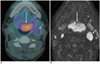
For an adequate approach, it is essential to identify the tumor regions with abnormal metabolic activity which can supply adequate materials for analysis. The development of new functional magnetic resonance imaging (MRI) techniques, including the DWI, improved the diagnostic yield which provides information regarding tissue cellularity and cellular membrane integrity for both primary lesion and nodal dissemination (Fig. 1). Nowadays, the DWI MRI can be used as an alternative tool when evaluating head and neck tumors (3). Erdem et al. (4) demonstrated that DWI was useful to differentiate benign from malignant thyroid nodules. In this study, ADC values were significantly lower in the malignant tissue when compared to the benign tissue.
The MRI has evolved significantly over the past years and improved the diagnostic yield of cervical lesions suspicious for malignancy. In the modern concept of target therapy, DWI seems to be an advantageous alternative for imaging-guided biopsy. The identification of the most suspicious areas or lesions to be biopsied is rather important for therapeutic planning and response evaluation.

Figures and Tables

Fig. 1
Forty nine-year-old man with mass in base of tongue has been questioned for malignancy.
A. Axial FDG PET-CT image shows mass with intense FDG avidity (large arrow). Maximum SUV was high corresponding to 8.9 (not shown) and left cervical lymphadenopathy was found (small arrow). B. Axial DWI MRI showing hyper-intensity in tongue lesion (large arrow) with lower ADC level suspicious for malignancy corresponding to 0.8 × 10⁻³ (not shown) and lymphadenopathy (small arrow). Biopsy performed revealed squamous cell carcinoma.
kjr-14-697-g001

References

1. Wu EH, Chen YL, Wu YM, Huang YT, Wong HF, Ng SH. CT-guided core needle biopsy of deep suprahyoid head and neck lesions. Korean J Radiol. 2013; 14:299–306.
2. Sack MJ, Weber RS, Weinstein GS, Chalian AA, Nisenbaum HL, Yousem DM. Image-guided fine-needle aspiration of the head and neck: five years' experience. Arch Otolaryngol Head Neck Surg. 1998; 124:1155–1161.
3. Chawla S, Kim S, Wang S, Poptani H. Diffusion-weighted imaging in head and neck cancers. Future Oncol. 2009; 5:959–975.
4. Erdem G, Erdem T, Muammer H, Mutlu DY, Firat AK, Sahin I, et al. Diffusion-weighted images differentiate benign from malignant thyroid nodules. J Magn Reson Imaging. 2010; 31:94–100.
We appreciated the comments from Dr. Guimaraes (1) regarding our recent article: "CT-Guided Core Needle Biopsy of Deep Suprahyoid Head and Neck Lesions" (1). We thank the editor for giving us the opportunity to reiterate the clinical use of the CT-guided needle biopsy in post-treated head and neck cancer patients.
In post-treatment cancer patients, it is important to differentiate tumor recurrence from post-therapeutic changes when a mass is presented in the irradiated areas. Timely salvage treatment offers a chance to cure the disease, but may introduce serious treatment-related complications. Therefore, an accurate tissue diagnosis is essential before the initiation of any salvage treatment. As illustrated in our paper, a CT-guided core needle biopsy could offer definitive tissue diagnosis under such circumstances.
The role of diffusion-weighted (DW) imaging in the evaluation of head and neck tumors seems promising. It appears to be useful in tissue characterization, nodal staging, therapeutic monitoring, as well as in the early detection of treatment failure (2). A lesion with a lower apparent diffusion coefficient (ADCs) may suggest malignancy as compared with that of a higher one (2). However, given that there are heterogeneous groups of benign and malignant lesions in the head and neck, overlapping of their ADC values is expected. Furthermore, DW imaging may not be sensitive enough to detect small foci of viable cancers dispersed within a large post-treatment inflammatory mass. In clinical practice, a single ADC threshold cannot be applicable for every site and all pathologic processes. More improvements might be brought about by standardization of the parameter, including selection of the best b-value, conquering the susceptibility and motion artifact in the head and neck area, and improving the degraded image quality by failed fat suppression and signal loss (2, 3).
In our institute, image-guided needle biopsy is a well acceptable means for obtaining definitive diagnosis in head and neck cancer patients with suspicious deep-seated recurrence. However, we believe that DW imaging in the head and neck area may serve as a complementary tool to routine MR imaging in the prediction of tissue diagnosis and in the identification of the area of most suspicion prior to biopsy, provided that the technical challenges, as mentioned before, have been properly met.

References

1. Wu EH, Chen YL, Wu YM, Huang YT, Wong HF, Ng SH. CT-guided core needle biopsy of deep suprahyoid head and neck lesions. Korean J Radiol. 2013; 14:299–306.
2. Thoeny HC, De Keyzer F, King AD. Diffusion-weighted MR imaging in the head and neck. Radiology. 2012; 263:19–32.
3. Vandecaveye V, De Keyzer F, Dirix P, Lambrecht M, Nuyts S, Hermans R. Applications of diffusion-weighted magnetic resonance imaging in head and neck squamous cell carcinoma. Neuroradiology. 2010; 52:773–784.
TOOLS
Similar articles