

 PDF
PDF ePub
ePub Citation
 Print
Print


INTRODUCTION
Arterial closure devices are increasingly used following transfemoral cardiac and peripheral vascular interventions. They can achieve faster hemostasis after sheath removal and allow early ambulation of the patient and improve patient comfort (1).
The Angio-Seal is a widely used closure device. Although it has many benefits, femoral artery stenosis and occlusions may result in claudication or limb ischemia (2), though the incidence is low at 0-2.9% (3). In a currently published series, endovascular treatment for complications at the common femoral artery or the superficial femoral artery, with or without stent implantation, showed excellent immediate and long-term outcomes (4). However, occlusions or embolisms that develop in more peripheral areas, such as popliteal artery embolisms, are much less common, and the optimal therapy for these cases remains uncertain.
Here, we report a rare complication related to an arterial closure device and its effective interventional treatment.
CASE REPORT
A 47-year-old man underwent percutaneous coronary intervention (PCI) secondary to unstable angina. The right common femoral arterial puncture site was closed with an Angio-Seal. There was no definite immediate complication, and the patient was discharged from the hospital.
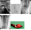
Two days after the PCI, he presented pain and numbness of the right calf. Symptoms were progressively aggravated in the following week. He revisited the hospital eight days after the procedure. Upon physical examination, there was no evidence of hematoma or pseudoaneurysm at the puncture site. Ultrasonography showed presumed aggregated collagen with bright echogenic anchor of the Angio-Seal device in the right popliteal artery (Fig. 1A).
A left femoral artery puncture was made, and right lower extremity angiography was performed with a 5 Fr catheter. Total occlusion of the distal popliteal artery was observed in angiography (Fig. 1B). Correlating with ultrasonography, a right popliteal artery occlusion due to the Angio-Seal that had broken away from its original location was diagnosed. The arteries distal to the occlusion were reconstituted by collateral vessels, showing reduced but persistent lumens.
Under local anesthesia, the right common femoral artery was exposed by a cardiothoracic surgeon. An 8 Fr long vascular sheath was inserted antegrade into the right superficial femoral artery. A 4 Fr Fogarty balloon catheter (Edwards Lifesciences, Irvine, CA, USA) was introduced into the vascular sheath, then inserted into the arterial lumen until the balloon was located distal to the embolus over an 0.018 wire (Terumo, Tokyo, Japan). The balloon of the Fogarty catheter was inflated and pulled back toward the femoral artery cutdown site (Fig. 1C). While performing the procedure, we could recognize the location of the embolus in angiography, which was seen as a filling defect between the balloon. Contrast was made by injecting a small amount of contrast material through the sheath (Fig. 1D). The embolus was successfully removed through the incision site of the femoral artery. It consisted of the Angio-Seal sponge with anchor, and had accumulation of acute thrombus proximally (Fig. 1E).
After a 12-month follow-up, the patient was pain free, and his post-interventional course was uneventful.
DISCUSSION
The Angio-Seal is a femoral arterial closure device which consists of an anchor composed of a polylactide and polyglycolide polymer, a collagen plug, and sutures contained within a special carrier system. The device achieves hemostasis by mechanically compressing the arterial puncture site between the anchor and the collagen plug (5, 6).
Various vascular complications from arterial closure devices are known, including hematoma, bleeding, arteriovenous fistula, pseudoaneurysm, arterial occlusion and infection (7, 8). Arterial stenosis and occlusions resulting in claudication or limb ischemia have been reported following Angio-Seal use in previous studies (2, 9).
In laboratory and clinical studies, it was found that when the Angio-Seal anchor is deployed in the artery, it is reabsorbed with complete dissolution in approximately 30 days as observed by microscopy, and in 90 days observed by chemical analysis (10). However, the intra-arterially-inserted sponge is unpredictable in terms of the extent and time course of bioabsorbability. Furthermore, a thrombus may accumulate while the sponge is dissolving, resulting in an increased size of the occlusive lesion and making complete extraction more difficult. Therefore, early removal of the occlusive materials is more suitable in comparison with conservative treatment (11).
The incidence of distal arterial embolism following Angio-Seal closure device deployment is more uncommon than proximal arterial occlusion (2, 9, 12). In review of the literature, only one case of popliteal artery embolism caused by Angio-Seal closure device was published (11). A goose neck snare was used to remove the embolic material in this report, but the procedure was incomplete. Fragmented parts of the suture and sponge were removed with a catheter. The patient experienced recurrent symptoms 6 months after the percutaneous embolectomy. In follow-up duplex sonography, a high-grade stenosis was noted in the tibiofibular trunk, and the patient required subsequent procedures.
The fluoroscopy-guided Fogarty embolectomy used in the present case did not require fragmentation of the embolus, but involved removal of the whole embolus by pulling back with an inflated balloon. The procedure can prevent not only the recurrence of stenosis, but also embolism of more distal arteries. Another advantage is that fluoroscopy provides clear visualization of the exact location of the embolus during the intervention when used with injection of small amounts of contrast material through the vascular sheath. It is a more focused and minimally invasive procedure compared to conventional surgical repair or other endovascular techniques.
As arterial closure devices are increasingly used, the occurrence of various ischemic complications may be expected to increase in the future, and different therapeutic options for the proper treatment of such complications are needed. In our case report, the complication of distal artery occlusion caused by Angio-Seal embolism was effectively treated with fluoroscopy-guided Fogarty embolectomy. We believe this new approach is appropriate to treat this type of complication, as it is a simple, safe, and accurate technique.
 XML Download