

 PDF
PDF ePub
ePub Citation
 Print
Print


INTRODUCTION
Pulmonary cryptococcosis is a type of fungal infection, initiated by inhalation of the organism, Cryptococcus neoformans, from an environmental source (1). It most commonly occurs in immunocompromised hosts, such as human immunodeficiency virus (HIV)-infected or cancer patients, and rarely involves the immunocompetent hosts (1, 2). Imaging features, associated with pulmonary cryptococcosis, which mimic hematogeneous metastases, are rare in immunocompetent patients. To our knowledge, this is a rare presentation of pulmonary cryptococcosis with computed tomography (CT) characteristics of hematogeneous lung metastases, and it's valuable to show the pathologic correlation with positron emission tomography (PET) scan.
CASE REPORT
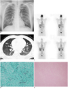
A 49-year-old man was admitted with a complaint of dry cough over the last two weeks, and pulmonary nodules were incidentally found on his chest radiography (Fig. 1A) at our outpatient department. Due to the pulmonary nodules of unknown cause, CT of the chest was arranged. Multiple well-defined nodules (number: more than 10) were observed in both lungs (Fig. 1B). Each nodule differed in size, ranging from 4 mm to 12 mm; moreover, the nodules were randomly distributed in every lobe, accompanied by minimal pleural effusions.
The patient denied being easily fatigued or having night sweats or rapid weight loss. No recent travel history was noted. The physical examination and all the laboratory tests presented no remarkable findings. We initially considered the nodules to have arisen from hematogeneous metastases. However, tumor markers, such as carcinoembryonic antigen, squamous cell carcinoma antigen, and prostate-specific antigen were all detected to be in the normal range. The gastroscopy and colonoscopy also showed within normal findings. After failing to find any possible primary malignancy, we arranged a whole body 18F-fluorodeoxyglucose (FDG) PET scan to evaluate the possible primary malignancy. The whole body FDG PET scan revealed no abnormal FDG uptake (Fig. 1C). Our preoperative diagnosis of hematogeneous lung metastases was based on the presence of multiple pulmonary nodules of varying sizes, which distributed randomly throughout the lung; however, the findings of the whole-body FDG PET scan indicated that the nodules were possibly benign.
Two nodules each from the right middle and lower lobe were surgically removed; Histopathology revealed a fibrotic nodule with mild peripheral granulomatous inflammation and many cryptococcal spores (Fig. 1D) within the central necrotic area (Fig. 1E). No evidence of malignancy was noted.
After diagnosing pulmonary cryptococcosis, a series of examinations were performed for greater detail, such as immunoglobulin levels, HIV by enzyme-linked immunosorbent assay, serum and cerebrospinal fluid cryptococcal antigens, but tested negative. The patient was then administered intravenous fluconazole, shifting to oral drugs 400 mg per day, for three months, after discharge. The nodules remain stable in size and number, during a one year follow up.
DISCUSSION
The pulmonary manifestations in patients with cryptococcal infection can present with a variety of appearances, showing from air-space consolidation to ill- or well-defined nodules/masses (2-7). In some cases, mixed associated findings, such as mediastinal lymphadenopathy, pleural effusions or cavitation, may be also noted. The number of nodules was often less in immunocompetent patients than in the immunocompromised ones (6, 7) In our patient, CT scans revealed multiple nodules, demonstrating relatively aggressive characteristics. The varying sizes of the pulmonary nodules and the random bilateral distribution mimicked the presentation of hematogeneous pulmonary metastases. Further, the nodular margins were well-defined without other associated findings, which was difficult to link to an infectious process.
On reviewing recent case studies and articles (Table 1), we noted that most multiple cryptococcal nodules have a tendency to cluster in one or few lobes in the immunocompetent patients, but cryptococcosis may also rarely show scattered nodules, such as in our case. Besides the aggressive nodular pattern, the totally absence of FDG uptake is also rare. There are only few studies combining the CT pattern showing pulmonary nodules with the discussion of PET signals. The standardized uptake value (SUV) of the cryptococcosis in these reports ranged from 0.93 to 11.6 (7, 9). Some lesions may be interpreted as benign by showing low SUVs. But most cases are interpreted as likely malignant lesions before biopsy because of the high SUV (9).
In our patient, PET scan provided functional information regarding the pulmonary lesions. A SUV of 2.5 has been traditionally used as a cut-off value for differentiating malignancy (7), but in pulmonary cryptococcosis, the SUV may vary widely, from mild to marked uptake (7, 9). In our patient, the intensity of FDG uptake was virtually absent for each lung lesion, which may indicate a low probability of malignancy (10). But FDG uptake generally depends on the size of the nodule. The small size (4-12 mm) of nodules, in this case, might also contribute to the absence of FDG uptake; hence, we decided to perform an open lung biopsy. Histopathologic results explained the cause of absence of FDG uptake: the nodule mainly composed of fibrotic and necrotic changes, with mild inflammatory cell infiltration. The lack of active inflammatory process told us the reason for absence of FDG PET signals.
In conclusion, we report an image presentation of pulmonary cryptococosis, which mimics hematogeneous metastases. PET did not demonstrate FDG uptake, suggesting a low probability for malignancy, and it correlated well with the pathologic finding: an infectious nodule composed of fibrosis and necrosis. It might be a crucial piece of information for clinicians when making management decisions, in cases of multiple lung nodules mimicking hematogeneous metastasis.

 XML Download