

 PDF
PDF ePub
ePub Citation
 Print
Print


INTRODUCTION
When we encounter an abnormal imaging finding of diffuse intense fluorodeoxyglucose (FDG) uptake in the unilateral breast, we usually take into account of the malignant conditions first, such as advanced breast cancer, breast lymphoma, or metastasis to breast. However, intense FDG uptake in the unilateral breast can be demonstrated by atypical breastfeeding practice. We report an interesting case of unusual unilateral breast FDG uptake in a recently lactating patient, due to the practice of breast-feeding only from her left breast.
CASE REPORT
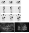
A 38-year-old woman underwent 18F FDG positron emission tomography (PET) for a follow-up after thyroidectomy. Three years ago, she was diagnosed as papillary thyroid cancer and underwent total thryoidectomy and modified radical neck dissection. After the operation, she received radioablation therapy. On a whole body FDG PET, diffuse intense FDG uptake was demonstrated in the entire left breast (Fig. 1A-C). There was no abnormal FDG uptake in other organs. On clinical examination, the breasts were soft without lumps.
Mammography and breast ultrasound were performed for further evaluation of the left breast. There was no mass or abnormal finding in the breast ultrasound (Fig. 1D). On mammography, her left breast was asymmetrically larger than the right breast without mass or focal asymmetry (Fig. 1E). She had delivered the baby 14 months before and stopped breast-feeding 1 day before the FDG PET. Her one-year-old infant had refused to feed from the right breast and she fed only from her left breast. Intense FDG uptake in her left breast was likely caused by normal physiologic lactation and no additional work up was required.
DISCUSSION
Recently, FDG PET has become an essential tool for cancer detection, staging and restaging of multiple malignant neoplasms. However, FDG is not a tumor-specific probe. It can accumulate in non-neoplastic conditions, including infection, postoperative scar, postradiation therapy and breast-related activity, in addition to its normal physiologic accumulation (1). Increased FDG uptake in lactation is thought to be the result of uptake of plasma glucose for milk production and increased activity of the ductal muscles. Significant numbers of women breast-feed their babies from one breast only, or more from one breast than the other from various reasons, causing considerable asymmetry of the breast. Hicks et al. (2) described that most of patients who were breast-feeding an infant before PET scanning had diffuse and symmetric uptake in the breasts. However, there was no breast uptake in the woman who had not begun breast-feeding without receiving medication to reduce prolactin secretion. Accordingly, intense uptake of FDG in lactating breasts appears to be related to suckling rather than prolactin. In addition, they also reported that high uptake and retention of FDG in the breast even after expression of breast milk suggests intracellular trapping of radiotracer in active glandular tissue. Consequently, increased radiation exposure to breastfeeding infant occurs mainly due to close contact to persons injected with FDG, rather than excreted milk.
Clinically, differential diagnoses of diffuse intense FDG uptake in the unilateral breast include advanced breast cancer, breast lymphoma and inflammatory condition. Mammography and ultrasound can be helpful in the differential diagnosis. When it comes to advanced breast cancer, mammography and ultrasound demonstrate abnormal mass, distortions and pathologic enlarged lymph nodes. Primary breast lymphoma of the breast is a rare disease accounting for 0.05-0.53% of all breast malignancies (3). The radiologic features of breast lymphoma are not pathognomic, presenting a wide spectrum of appearances, ranging from solitary or multiple well-defined to ill-defined masses to diffuse trabecular and skin thickenings (4). Infectious disease of breast usually shows characteristic clinical presentation, as well as typical imaging features, such as diffuse skin thickening, increased echogenicities, and irregular hypoechoic mass.
For the accurate interpretation of FDG PET findings, we should understand the normal physiologic uptake of tracer and benign pathologic conditions, which can be mistaken for malignancy (5). In our case, before we knew about the history of her breastfeeding, we had considered abnormal FDG PET findings as an advanced breast cancer. However, there were no abnormal findings on mammography and ultrasound, and on questioning, the patient gave a history of breastfeeding only from her left breast.
In conclusion, when the diffuse intense FDG uptake is noted at the unilateral breast, and no abnormal lesion is observed on mammography and ultrasound, we should consider that they can be related to the practice of unilateral breastfeeding.
 XML Download