

 PDF
PDF ePub
ePub Citation
 Print
Print


INTRODUCTION
A pulmonary mucinous cystadenocarcinoma (PMC) is an extremely rare tumor that has recently been considered to be part of the colloid adenocarcinoma spectrum. We present a case of a PMC in a 54-year-old woman along with a literature review focusing on the characteristic CT features that may help differentiate between PMCs and other mucinous or cystic lung lesions.
CASE REPORT
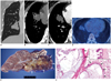
A 54-year-old asymptomatic woman was found to have a mass in the right middle lung zone on a routine chest radiograph. The results of the clinical examinations and routine laboratory tests were unremarkable. A chest CT showed a lobulated hypodense mass (4.3 cm maximum diameter) with peripheral stippled calcifications. The mass had well-defined margins, with no enhancement following contrast administration (5.7 Hounsfield unit) and had no hilar or mediastinal lymph node enlargement (Fig. 1A-D). 18F-fluorodeoxyglucose (FDG) positron emission tomography/computed tomography revealed mild heterogeneous FDG uptake (SUV = 2.6) (Fig. 1D). Initially, the lesion was diagnosed as adenocarcinoma with mucin production or mucinous adenocarcinoma by transbronchial lung biopsy. A right middle lobectomy was performed, and the specimen contained a lobulated, cystic, and gelatinous mass measuring 4.5 × 3.5 × 2.5 cm (Fig. 1E, F). Histopathologically, the tumor consisted of abundant mucin-filling cysts surrounded by thick fibrous walls. Columnar mucinous epithelial cells lined the thickened alveolar walls. Over the 9-month follow-up period, the patient remained free of recurrent disease.
DISCUSSION
A PMC is an extremely rare subtype of pulmonary adenocarcinoma that was first described by Gowar in 1978 (1). A PMC was considered a distinctive variant of adenocarcinoma by the latest edition of the World Health Oraganiztion classification (2). However, the 2010 International Association for the Study of Lung Cancer/American Thoracic classification system for lung tumors (2) includes PMC as part of colloid adenocarcinomas. This system suggests that these adenocarcinomas, consisting of uni- or oligolocular cystic structures by imaging and/or gross examination, be included in the category of colloid adenocarcinoma, and a comment could be made that the tumor resembles the formerly classified PMC. Some experts have included these tumors as distinct variants among a spectrum of mucus-producing adenocarcinomas, including mucinous adenocarcinomas, mucoepidermoid carcinomas, signet ring cell adenocarcinomas, mucinous cystadenocarcinomas, and mucinous "colloid" adenocarcinomas (3). A PMC is a cystic adenocarcinoma with copious mucin production and resembles the tumors of the same name in the ovary, breast and pancreas (4). According to Iwasaki et al. (5), 20 cases of PMC had been reported so far, in patients ranging from 29 to 75 years (median, 64 years) of age, with no patient gender difference (6). The majority of patients were asymptomatic and the lesions were discovered incidentally, but some presented with non-specific respiratory symptoms including cough, chest pain, dyspnea, and bloody sputum (5, 7, 8).
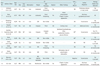
We screened the English literature and found 10 cases of PMC in which CT images of the disease were described (summarized in Table 1). Typically, PMC appears as a well-defined, partly-lobulated, homogeneous, and persistent-low attenuation lesion with focal mural or septal enhancement on CT (1, 9). In several cases (8, 9), as in ours, stippled calcifications were noted in the wall or septae of the lesion.
Although sometimes observed as unilocular cystic or low-attenuation lesions, most PMCs were shown as multi-locular cystic or multilobulated low attenuation lesions with septal or wall enhancement. However, PMCs may present with no septal or mural enhancement, which was the case in the present patient and in the report by Raza et al. (9). The problem is that we may have some difficulty in making a correct diagnosis of PMC as a neoplastic condition because it often shows bland radiologic findings such as a well-defined margin or absence of enhancement.
In the case of PMCs, FDG PET might show little FDT uptake, hence the tumor has low tumor cellularity and abundant mucin (10). On the other hand, Iwasaki et al. (5) reported a case which showed intense FDG uptake (maximum standardized uptake value, 6.7) in the solid portion of invasive carcinoma in the wall of the lesion. Similar findings were also reported by Raza et al. (9), who found that the enhancing portion of the mass showed FDG uptake. Considered altogether, because enhancing thick wall is probable cancer foci, we suggest that FDG uptake of PMC may depend on the thickness of the wall. Therefore, FDG PET may not be enough to exclude the possibility of malignancy such as PMC, especially in tumors without visible thick and enhancing walls.
Differential diagnosis of PMC should include the broad spectrum of disease entities such as mucin-producing lung neoplasms (mucinous ["colloid"]), carcinomas and mucinous adenocarcinomas, metastatic mucinous carcinoma and mucinous/mucinous-looking non-neoplasms (mucocele and bronchogenic cyst), as well as lymphangiomas (8, 9). Cytologically, mucinous carcinomas may resemble that of PMCs; however, they usually lack the cystic changes and a fibrous capsule, so they appear to be solid rather than cystic on radiography (1, 5). However, common radiologic features of mucinous carcinoma consist of an ill-defined, solid peripheral nodule or lobar consolidation, and ground-glass opacity (11). Differentiating PMC from mucinous adenocarcinoma on pathologic evaluation may often be a challenge, especially in those unusual cases where necrosis results in secondary cavitation or cyst formation in the latter (12). However, because they show distinct features on CT, CT will help distinguish between the two. Namely, mucinous adenocarcinoma is diffuse and often bilateral, showing grossly as an ill-defined lobar consolidation, while a PMC usually presents as a well-circumscribed low-attenuating lesion (12-14). The well-defined border of the PMC may be related to the histologic features of the tumor in which thick fibrous walls surround abundant mucin filled cysts. Solitary metastatic mucinous tumors from other organs, especially from the ovary, breast, and pancreas, may mimic primary PMC, therefore, thorough clinical and radiologic evaluation is mandatory to exclude metastases from other organs (15). Furthermore, PMCs may present similarly with mucoceles/bronchoceles, and we should consider PMCs as differential diagnoses in patients presenting with the gloved-finger appearance of a mucocele that has suspicious imaging features including focal thickening or enhancement of the walls and septa on enhanced CT scans (9).
Preoperative diagnosis is very difficult to establish, mainly because of the composition of the tumor (i.e., large amount of mucin and small number of malignant cells) (1, 8, 15). Only two cases were preoperatively diagnosed by using fine needle aspiration cytology (16, 17). Maeda et al. (13) described the invasive preoperative diagnostic procedures including transbronchial lung biopsy and transthoracic needle biopsy as being generally non-diagnostic because of the composition of the tumor.
The biological behavior of PMC is generally unknown, due to the limited number of reported cases. However, PMC appears to have a low malignancy potential, although it is considered malignant (8). The tumor seems to have a favorable clinical course when compared with other primary bronchogenic carcinomas (1); neither distant metastasis nor recurrence has been reported (15). Therefore, a formal anatomic lobectomy for early stage tumors is probably the correct surgical approach as mentioned by Mann et al. (19). The role of adjuvant therapy chemotherapy or radiotherapy is not established (7), and given the unclear natural history of PMC, careful long-term follow-up study is probably needed.
In conclusion, a mucinous cystadenocarcinoma is a rare malignant neoplasm which should be included among the differential diagnoses of mucinous lung neoplasms or mucinous cystic lung lesions. The awareness of theses imaging features including a well-defined, partly-lobulated, homogeneous, and low-attenuated lesion with focal mural or septal enhancement on CT with variable FDG uptake on PET, may allow us to include the PMC as a probable differential diagnosis among lung tumors appearing as low-attenuation soft-tissue mass on CT with variable FDG uptake on PET.
 XML Download