

 PDF
PDF ePub
ePub Citation
 Print
Print


INTRODUCTION
Angiomyxolipoma is a rare variant of lipoma, characterized by the proliferation of mature adipose tissue associated with myxoid stroma and multiple vascular structures. To date, 11 cases of angiomyxolipoma have been reported in the literature. Among them, angiomyxolipoma of the spermatic cord has only been reported once, by pathologist Mai et al. (1) in 1996, and this report focused on histology. Here, we report for the first time, the imaging findings of angiomyxolipoma of the spermatic cord in an elderly man which presented initially as an inguinal hernia.
CASE REPORT
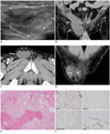
A 72-year-old man presented with a 1 month history of a painless palpable mass in the right inguinal region. On physical examination, an irreducible tender mass was detected in the right inguinal region. Laboratory data were within normal ranges. A clinical diagnosis was made of a right inguinal hernia. The inguinal US revealed an oval shaped, mixed echogenic mass (Fig. 1A) with a focal area of slightly increased vascularity on a color Doppler image. A longitudinal scan showed a hyperechoic area on the superficial one-fifth region, a linear structure on the intervening portion, and a mixed echoic area on the deep four-fifth region of the mass (Fig. 1A). There was no evidence of a herniated bowel loop or omental fat. Contrast enhanced multi-detector (MD) CT demonstrated an approximately 9.8 × 5.3 cm well-defined elongated, homogeneously low attenuated mass without an enhancing portion attached to the right spermatic cord and extending to the right inguinal subcutaneous region (Fig. 1B, D). The axial image showed the focal fat density area without an enhancing portion, on the anterior peripheral portion of the mass (Fig. 1C). There was no radiographic evidence of any regional lymph node involvement or invasion involving a contiguous structure on CT scan. Preoperative diagnosis of benign soft tissue tumor of the spermatic cord was suggested because the imaging appearances were not compatible with inguinal hernia or inguinal hydrocele.
Upon inguinal exploration for this suspected soft tissue mass, a 10 × 6 cm soft gelatinous mass was discovered which was adherent to the spermatic cord but simultaneously mobile and free from attachments with other soft tissue elements. The spermatic cord coursed through this mass. Upon closer inspection there was no evidence of a hernia or laxity in the posterior wall of the inguinal canal. The patient underwent excision of the mass without hernia repair surgery.
The histopathologic findings showed a well-demarcated, myxoid neoplasm comprised of spindle and stellate cells, mixed with mature adipocytes and numerous thin- and thick-walled vessels of various sizes (Fig. 1E). Immunohistochemically, the cells of the myxoid areas were positive for CD34 (Fig. 1F) but not for S-100 protein, desmin, or smooth muscle actin (SMA). The mature adipocytes were focally positive for S-100 protein (Fig. 1F). The CD34 and SMA highlighted vascular endothelial cells and perivascular smooth muscle fibers, respectively (Fig. 1F). The tumor was negative for desmin (Fig. 1F). On the basis of these findings, angiomyxolipoma was diagnosed.
DISCUSSION
Spermatic cord tumors are usually benign lipomas discovered incidentally during inguinal herniorrhaphy (2). Some myxoid type spermatic cord tumors that have been reported are thought to have been diagnosed as inguinal hernia on clinical examination (2-4). However, variable myxoid tumors of the spermatic cord, including angiomyxolipoma, aggressive angiomyxoma, myxoid liposarcoma, intramuscular myxoma, neurofibroma with myxoid change, myxoid or spindle-cell lipoma, and superficial angiomyxoma have a non-specific imaging appearance (4). Aggressive angiomyxoma appears hypoechoic on US, and are usually hypodense on CT (5). Kim et al. (6) suggested that myxoid components of liposarcoma in four patients showed predominant regions in which CT attenuation was less than that of muscle and similar to that of water before contrast enhancement. Because of these non-specific imaging appearances, preoperative radiological diagnosis of myxoid tumor of the spermatic cord can be difficult in daily practice.
Angiomyxolipoma is a rare form of lipoma. Angiomyxolipoma shows a characteristic gross appearance of a gelatinous, beige-yellow cut surface. The histopathologic features of angiomyxolipoma are characteristic and include an admixture of paucicellular myxoid areas and mature adipocytic areas without lipoblasts, and both components contain numerous thin- and thick-walled vessels. In myxoid areas, spindle cells are positive for CD34 and vimentin expression, and negative for SMA, desmin, and S100 protein expression (1, 7, 8).
While 11 cases of angiomyxolipoma have been reported in the literature, there has been only one report of angiomyxolipoma arising from the spermatic cord (1, 7, 8). The case report was published in 1996 and contained histopathologic findings of angiomyxolipoma of the spermatic cord, but the imaging appearance of angiomyxolipoma of the spermatic cord remained an unreported entity. To the author's knowledge, only one previous report has demonstrated the US and magnetic resonance imaging (MRI) appearance of angiomyxolipoma in the knee.
Angiomyxolipoma has been described by Kim et al. (8) as appearing on US as a well-defined mixed echoic mass with an area of increased vascularity on a color Doppler image and on MRI as a heterogeneous signal intensity at all MR sequences. These findings were thought to be the result of a heterogeneous mixture of myxoid, adipose tissue, and vascular components (8). Our case presented as a mixed echogenic mass on US. A hyperechoic area (due to fat component) was seen in a superficial area, and a mixed echoic area (due to myxoid component) was demonstrated on US. The spermatic cord was observed traversing between the two areas on both US and CT scans (9). These imaging findings may help to determine the radiological diagnosis of spermatic cord tumor rather than inguinal hernia. CT demonstrated a well-defined, homogeneously low density mass which contained a focal peripheral fat density area. No contrast enhancement was registered. In retrospect, the attenuation value for the homogeneous mass portion (except the focal fat density area) was measured as +16 Hounsfield units (HUs). According to the literature, 17 HU with a non-enhanced appearance of the mass is thought to be the result of a myxoid component of a gelatinous gross appearance of the tumor (10).
For the patient with an irreducible inguinal mass, it is important to make a prudent preoperative radiological diagnosis with a high suspicion for spermatic cord tumor in order to perform the proper therapeutic strategy. Because of its rarity, angiomyxolipoma of the spermatic cord is often not considered in the differential diagnosis of an inguinal hernia. However, angiomyxolipoma should be considered in the differential diagnosis in patients with an inguinal mass, while imaging diagnosis, such as US, and CT may help in making a preoperative diagnosis.
In conclusion, an angiomyxolipoma of the spermatic cord should be considered in the differential diagnosis in patients with an inguinal mass with a myxoid component containing a fat portion in the inguinal region on CT scan.
 XML Download