

 PDF
PDF ePub
ePub Citation
 Print
Print


INTRODUCTION
Follicular dendritic cell sarcomas were first described by Monda et al. (1) and are known to occur predominantly in adults, though they can occur at any age. This tumor arises from germinal centers and mainly affects lymph nodes. Extranodal sites include the palate, tonsils, tongue, intra-abdominal soft tissue, liver and spleen (2). Only a few reports have discussed the pathology and none have discussed the CT findings of this tumor when occurring in the greater omentum (3).
We present the CT findings from a 47-year-old female patient with a follicular dendritic cell sarcoma of the greater omentum.
CASE REPORT
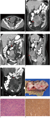
A 47-year-old woman had had epigastralgia, acid regurgitation, and fatigue over the two prior months. Her body weight had increased 10 kg over one month and a physical examination revealed a large, bulging, movable hard mass in the umbilical area. No bowel obstruction, anorexia, fever, or tarry stool was noted. A colonoscopy showed hyperplastic polyps, while a gastroduodenoscopy revealed a gastric polyp and erosive gastritis. CT of the abdomen showed a 14 × 8 × 7-cm mass with calcification in the abdomen and pelvis at the umbilical level (Fig. 1A, B). The mass was inferior to the stomach, anterior to the transverse colon, and extended downward to pelvic level (Fig. 1C, D). We could trace the engorged right gastroepiploic artery and vein at the uppermost part of the tumor (Fig. 1E). All of these landmarks revealed that the exact anatomical location was the greater omentum. The tumor showed complex internal architecture, such as heterogeneous enhancement, central necrosis, hemorrhage, and focal calcification. The possibility of sarcoma and primary gastrointestinal stromal tumor (GIST) were considered. Under the impression of sarcoma of the greater omentum or primary GIST of the greater omentum, an operation was performed.
The surgery revealed a huge solid encapsulated mass in the omentum, with central necrosis and hemorrhage within the tumor (Fig. 1F). No invasion to adjacent organs was observed and a histological diagnosis revealed a follicular dendritic cell sarcoma of the greater omentum (Fig. 1G, H).
DISCUSSION
A follicular dendritic cell sarcoma is a rare tumor arising from germinal centers and affecting mainly lymph nodes. It affects patients of any age, but mainly adults of either sex (4, 5). Extranodal sites that have been reported to include the palate, tonsils, tongue, intra-abdominal soft tissue (mesentery, mesocolon and greater omentum), stomach, small intestine, colon, pancreas, liver, spleen, mediastinum, retroperitoneum, intracranium, breast, abdominal wall, thyroid gland, lung and the dura mater of the spine (3, 5-7). The clinical symptoms and laboratory findings include low-grade fever, weight loss, mild anemia, and hypergammaglobulinemia. Intra-abdominal tumors have an aggressive course (4); follicular dendritic cells participate in the immune system by presenting antigens for B cells and by stimulating B-cell proliferation and differentiation (7). These tumors have distinct immunohistochemical features, with the diagnosis relying on the reactivity to CD21 and CD35 markers (8).
There are a few reports of the imaging features of follicular dendritic cell sarcoma arising in the abdomen (9, 10). The radiologic findings of follicular dendritic cell sarcoma in the greater omentum have not been described before, to our knowledge. Leipsic et al. (11) reported a case of mediastinal follicular dendritic cell sarcoma, described as coarse and chunk-like on CT images. Calcification also was demonstrated in our case. On the post-contrast images of our study, heterogeneous densities within the tumor were much more apparent; on histology, the enhancing portion corresponded to tumor and the non-enhancing area was focal necrosis and hemorrhage. The large tumor size of our case can be related to the presence of gross necrosis and hemorrhage; similar findings were noted by Long-Hua et al. (10).
The first issue in our case was to identify the exact anatomical location of the tumor. To understand the embryology and anatomy of the greater omentum is important for a radiologist. The greater omentum is composed of a double layer of peritoneum that extends from the greater curvature of the stomach inferiorly. After a distance that usually ranges from 14 to 36 cm, the greater omentum turns superiorly on itself to drape below the transverse colon. The normal greater omentum appears on CT scans as a band of fatty tissue of varied width (12). A multi-detector CT scan with multiplanar reformation allows for the determination of anatomical details of the omental mass, which is embedded in fatty tissue between the stomach and the colon. In our case, we could identify the mass by coronal plane imaging as below the stomach with no connection to it. The tumor was located anterior to the transverse colon, and draped to the pelvis. This was seen in coronal, axial, and sagittal planes. The other crucial point is to recognize the relationship between the gastroepiploic vessels and the mass, because the blood supply of the greater omentum is largely originates from the anastomosis of the right and left gastroepiploic arteries. The right gastroepiploic artery and vein were seen at the highest part of the tumor, with branches to the mass. Accurately locating the greater omentum leads to a correct differential diagnosis. Metastatic disease involving the greater omentum is far more common than primary tumors. However, the tumors are usually multiple small foci. A solitary soft tissue mass could be a primary neoplasm of the omentum. These most often arise from mesodermal elements and are malignant. Primary tumors include leiomyosarcoma, hemangiopericytoma, fibrosarcoma, reticulosarcoma, spindle cell sarcoma, liposarcoma, and primary GIST.
The omentum is composed of four layers of peritoneum and calcification may occur in several types of peritoneal tumors, such as the primary peritoneal serous carcinoma, desmoplastic small round cell tumor, mesothelioma, and follicular dendritic cell sarcoma. Calcification has been reported to occur in 30% of primary peritoneal serous carcinoma cases (13); however, most of these cases are diffuse neoplastic processes. Calcification within diffuse peritoneal malignant mesothelioma is considered rare. Desmoplastic small round cell tumor occurs predominantly in adolescents and young adult males. Primary GIST in the omentum and follicular dendritic cell sarcoma has overlapping appearances on imaging. Both of these two tumors are well-circumscribed large masses containing areas of low attenuation due to necrosis and hemorrhage. However, Kim et al. (14) reported no calcification in their cases of primary GIST in the omentum. The depiction of coarse and chunk-like calcification of an omentum tumor on CT images can raise the suspicion of a follicular dendritic cell sarcoma.
In summary, multi-detector CT is the best imaging modality to depict greater omentum lesions. The anatomic relationships of the soft tissue mass, stomach, colon, and the gastroepiploic vessels in multiple planes are very useful for diagnosis of omentum tumors. A follicular dendritic cell sarcoma should be considered in the differential diagnosis of a solitary omentum mass, especially one with coarse and chun-klike calcification.
 XML Download