

 PDF
PDF ePub
ePub Citation
 Print
Print


INTRODUCTION
A cholesterol granuloma consists of fibrous granulation tissue containing cholesterol crystals that are surrounded by foreign body giant cells; the occurrence of this disease in the breast is very rare. It is not difficult to distinguish a cholesterol granuloma from a breast carcinoma on histological examination; however, the clinical findings and radiographic images of the cholesterol granuloma are similar to those of cancer, leading to a possible confusion between these two diseases (1).
Few studies describe the imaging findings of cholesterol granuloma (1-4), and only one case has been reported in the English literature: a cholesterol granuloma presenting as an intracystic mass (5). We present a similar case, a cholesterol granuloma presenting as an intracystic mass, appearing to be a papillary neoplasm by ultrasonography. Enhanced characterization of this disease may help in distinguishing it from breast cancer on radiologic studies.
CASE REPORT
A 62-year-old woman with no previous history of breast disease was referred to our hospital from a local clinic. The patient complained of a painless, palpable mass in the lower, outer quadrant of her left breast; the mass had been present for 10 years. She exhibited no other clinical symptoms or other signs upon physical examination, and she had no history of having undergone a breast biopsy or chest wall trauma.
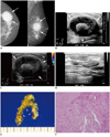
Mammography results revealed a well-circumscribed, round mass in the lower, outer quadrant of the left breast. The mass was generally of high density and had a peripheral radiolucent rim, with scattered coarse, heterogeneous calcifications present (Fig. 1A).
On ultrasonography, a 5 × 4.6 cm circumscribed, oval, complex echoic mass was observed in the left breast, at the 4 o'clock position (Fig. 1B). Within this cystic mass, there was a lobular, solid lesion of approximately 2 cm in diameter with floating, hyperechoic internal contents. No increased vascularity was visible on color Doppler imaging (Fig. 1C). The lesion met the criteria for Breast Imaging Reporting and Data system category 4a, and we therefore recommended a core needle biopsy.
Ultrasound-guided core needle biopsy of the mass was performed with a 14-gauge needle. The mass collapsed after biopsy, and the residual lesion measured 3.7 × 0.8 cm (Fig. 1D).
Although the mass in the left breast was confirmed to be benign, we performed a resection in order to alleviate the patient's symptoms. The resected tumor was determined to be a cholesterol granuloma. The surgical specimen measured 4.5 × 4.0 cm and was primarily composed of cystic material with internal brown, necrotic material (Fig. 1E). Microscopic examination revealed a thick, fibrous cystic wall, with characteristic cholesterol crystals arranged in parallel or radial arrays, and aggregates of histiocytes (Fig. 1F). Hemorrhage (both fresh and old) and fat necrosis were also noted.
DISCUSSION
Cholesterol granuloma commonly occurs in the middle ear cavity and in the mastoid process, but is very rarely observed in the breast. On histological examination of the granuloma mass, it was found to be a fibrous granulation tissue containing cholesterol crystals, surrounded by foreign body giant cells (1).
The pathogenesis of breast cholesterol granuloma is still controversial, and 2 theories have been suggested. One theory proposes that the primary disease is duct ectasia, with periductal inflammation causing the escape of fat-rich intraluminal material. The other theory purports that duct ectasia follows periductal inflammation with ductal wall damage. Irrespective of etiology, this disease is usually accompanied by duct ectasia and is thought to be a rare manifestation of that entity. The presence of fat-rich material resulting from duct ectasia causes cholesterol crystals to form; the crystals then infiltrate the granulomatous tissue and create a cholesterol granuloma (1, 4). A history of previous biopsy or trauma to the breast, sufficient to cause rupture of an ectatic duct, is associated with the formation of cholesterol granulomas (1). In this case, our patient had no history of breast trauma or breast biopsy, but mammary duct ectasia was visible on microscopic analysis of the biopsy specimen.
Breast cholesterol granuloma has multiple clinical manifestations. The most common finding is a painless, palpable mass; our patient complained of having this symptom for the 10 years prior to presenting to our hospital. Duration and stability of symptoms are helpful signs to allow clinicians to differentiate this type of mass from breast cancer.
The reported mammographic findings of cholesterol granulomas have been nonspecific, but calcification was observed in several cases (2). A few cases of cholesterol granuloma of the breast have been reported: in 5 cases, the ultrasonographic findings were described (1-5); in 4 cases, a solid nodule mimicked breast cancer (1-4); and in one case, the patient presented with an intracystic mass, similar to our case (5). Smith et al. (5) described a large cyst with a 2 cm papillomatous mass within the cyst. In our case, the cholesterol granuloma appeared as a 5 cm complex echoic mass with a 2 cm intracystic, solid lesion. There were multiple floating, hyperechoic specks within the cyst, and the intracystic lesion showed no increase of vascularity on color Doppler imaging.
Ganesan et al. (6) reported the ultrasonographic features of papillary neoplasm, such as the shape and vascularity of the intraductal lesion, and associated findings such as ductal dilatation. Papillary neoplasm usually appears as an intracystic solid lesion with peripheral fronding in the dilated duct, with secondary ductal dilatation proximal to the mass (6). Moreover, the intracystic solid lesion showed increased vascularity. These findings did not apply to our patient, and this likely helped the radiologists to differentiate the cholesterol granuloma from a papillary neoplasm.
Cholesterol granuloma of the breast is not suspected to be a risk factor for breast cancer, but the few reports in the literature thus far have recommended confirmation of the presence/absence of cancer after histological analysis of surgical excision or excisional biopsy specimen. Furuhira et al. (3) described a breast cholesterol granuloma accompanied by cancer. On ultrasonographic analysis, they observed a lobulated, heterogeneous mass composed of 2 parts: a hyperechoic mass with increased posterior echogenicity and a hypoechoic mass with posterior shadowing. The mass was confirmed to be a cholesterol granuloma on fine needle aspiration, but the resected mass comprised a cholesterol granuloma with accompanying invasive ductal carcinoma. A surgical biopsy was performed because of the suspicious ultrasonographic finding, and results of histological analysis of the specimen differed from the results of initial needle biopsy. A rigorous image review and accurate histological examination are necessary to avoid misdiagnosis.
We report a breast cholesterol granuloma presenting as an intracystic mass and mimicking a papillary neoplasm. The radiological and clinical findings of this disease are analogous to breast cancer in the reported literature and in the present case; therefore, the mass is presumed ocassionally to be malignant. Although the imaging findings of breast cholesterol granuloma and intraductal papilloma can overlap, we suggest improved characterization of breast cholesterol granuloma to successfully differentiate it from breast cancer.
 XML Download