

 PDF
PDF ePub
ePub Citation
 Print
Print


INTRODUCTION
A segmental testicular infarction (STI) is a rare cause of acute scrotum. The etiologic mechanism is largely considered idiopathic, but cases have been associated with hypercoagulability disorders, vasculitis, torsion, trauma, infection, and iatrogenic vascular injury (1, 2). A STI usually presents with acute scrotal pain and may resemble epididymoorchitis or torsion. Clinically, a STI presents with acute scrotal pain and is indistinguishable from other causes of scrotal pain.
Ultrasonography (US) is the imaging method of choice for scrotal diseases. Variable appearances on US have been described previously depending on the evolution of a STI over time (1-4). The major concern in those studies was that the differentiation between a STI and testicular neoplasm could be challenging (4). Shear-wave elastography (SWE) is a new imaging modality that estimates tissue stiffness in real time (5). It was previously used for the assessment of breast, prostate, liver and thyroid diseases (6). Few reports concerning the efficacy of elastography for scrotal mass assessment are available (7). Testicular cancers were found to be harder than the normal testicular parenchyma on elastography (7). Therefore, our assumption was that a testicular neoplasm would be stiffer than the normal testicular parenchyma on SWE. We are not aware on the use of SWE for diagnosing a STI. Herein, we report a case diagnosed to have a STI based on the SWE features and emphasized its role in the differential diagnosis.
CASE REPORT
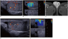
A 35-year-old man presented with left sided acute scrotal pain. He was previously healthy without any hematological disorder and had no prior trauma to the scrotum. He had no fever, and a urinalysis, serum electrolytes and blood count were normal. Clinical examination revealed tenderness of the upper pole of the testis. US examination was performed with a 4-15 MHz linear transducer (SuperSonic Imagine, Aix en Provence, France). An almost round inconspicuous isohyperechoic area was seen at the upper pole of the left testis on gray-scale US and color and power Doppler US revealed absent flow in this area (Fig. 1A). The remaining part of the left testicle and the right testis had a homogenous appearance. SWE was performed and showed soft areas on color mode in the central portion of this area with a mean elasticity value of 1.7 kPa (Fig. 1B). The normal testicular parenchyma had an elasticity value of 2.6 kPa. Gadolinium-enhanced magnetic resonance imaging of the testis revealed an avascular zone at the upper periphery of the left testis with perilesional hypervascularity (Fig. 1C). The tumor markers, including β human chorionic gonadotropin (<1.20 mIU/mL) and α fetoprotein (4.53 IU/mL), were unremarkable. A STI was the presumed diagnosis and a follow-up ultrasonography performed 10 days after the initial examination revealed that the size of the lesion decreased with an ill-defined wedge shaped hypoechoic appearance. The SWE showed a wedge shaped stiff area corresponding to a testicular lobule. SWE at the control examination showed an increased elasticity value (mean, 22.3 kPa) compared to the previous examination (Fig. 1D). Based on the US, color Doppler US and SWE findings, a STI was diagnosed.
DISCUSSION
The spectrum of findings on gray-scale and color Doppler US imaging of a STI differs depending on the time between the onset of testicular pain and the US examination (1, 3, 8). It was shown that lesions identified earlier were more often rounded and nearly isoechoic to the testis and have ill-defined margins (8). Based on the gray-scale US features at an early presentation, differentiation from a testicular neoplasm is difficult. Interestingly, we found a softer area compared to the normal testicular parenchyma at the initial SWE examination (within 24 hours after onset of pain). Increase in water content and swelling of the tissues secondary to ischemia may be the pathophysiological mechanism for the infarcted area that appeared soft on SWE led us to follow the patient rather than to go to surgery.
On gray-scale US, a STI became more conspicuous and smaller over time, hypoechoic to testis, and often having a wedge shape (8). A follow-up of our case showed that the lesion became wedge-shaped but still had ill-defined margins. On SWE, the lesion appeared stiffer than the testicular parenchyma, probably due to the organization of hemorrhage and necrosis and the shrinkage in size of the lesion. Interestingly, the lesion had the shape of a testicular lobule on SWE images that was more easily appreciated than the gray-scale US, enabling us to confidently diagnose a STI.
On color Doppler US, a typical STI has complete loss of vascularity; however, diminished vascularity has also been reported (1). Presence of color Doppler signal within the lesion results in diagnostic difficulties, with a tumour being a possibility (4, 9). Testicular tumours are normally vascular on color Doppler ultrasonography and may demonstrate a characteristic pattern of vascular flow (10). The demonstration of vascularity in a focal testicular mass is thought to be dependent on the size of the abnormality. Horstman et al. (10) failed to demonstrate an increase in lesion vascularity when the lesion was smaller than 16 mm in diameter. However, with modern ultrasonography equipment, color Doppler flow may be demonstrated in focal, solid lesions as small as 5 mm (11). In our case, at the initial examination, there was complete loss of vascularity; however, on follow-up Doppler US, there was some vascularity within the lesion. In our case, without knowing the previous Doppler US findings, the presence of vascularity within the lesion at follow-up may cause difficulties for the diagnosis.
Contrast-enhanced US was successfully used to improve lesion conspicuity and depict the anatomic characteristics in a STI (8). Infarction presents as one or more avascular areas separated by normal vessels at contrast-enhanced US, which is consistent with ischemic testicular lobules. The perilesional rim enhancement is a useful finding frequently seen in patients with a STI on contrast-enhanced US (8). A more pronounced perilesional rim enhancement and lobular distribution differentiates STI from an abscess. Similar to contrast enhanced US, gadolinium-enhanced magnetic resonance imaging demonstrates an enhanced halo delimiting the avascular area in a STI (3).
In summary, similar to gray-scale and Doppler US, the SWE features may depend on the evolution of a STI that is soft at initial presentation and hard the late stages compared to normal testicular parenchyma. The use of SWE in addition to gray-scale and Doppler US may prevent unnecessary orchiectomies.
 XML Download