

 PDF
PDF ePub
ePub Citation
 Print
Print


INTRODUCTION
Computed tomography coronary angiography (CTCA) is a non-invasive tool with high diagnostic accuracy for detection of coronary arterial stenosis (1-6). In retrospectively electrocardiogram (ECG)-gated spiral data acquisition mode (spiral mode), radiation exposure is high and radiation doses can be as high as 12 mSv (7). With prospective ECG-triggering axial acquisition mode (sequential mode, also referred to as "step and shot acquisition"), the average effective radiation dose can be as low as 2.1 mSv. For the first generation of dual-source CT (DSCT), the effective radiation doses of CTCA can be lowered to 1.5-2.5 mSv using prospective ECG-triggering axial acquisition mode (8).
The second generation of dual-source 128-slice CT systems (Definition Flash, Forchheim, Germany) provides a wide coverage of 38.4 mm, allowing CTCA examinations to be performed at a high-pitch, up to 3.4 (9). A gantry rotation time of 0.28 s enables a temporal resolution of 75 ms. Thus all axial planes that comprise the heart can be acquired in approximately 280 ms, and the CTCA acquisition time is reduced to a quarter of a second, depicting of the entire heart within a single heart beat. Previous studies indicate that prospectively ECG-gated high-pitch spiral acquisition mode (flash mode) CTCA provides a high diagnostic accuracy for the assessment of coronary stenosis with an effective radiation dose below 1 mSv (10, 11). This technique is limited by the need for regular sinus rhythm and low heart rates (HRs) (below 60-65 beats per minute, bpm) to ensure an adequate examination using the high-pitch spiral acquisition mode (11, 12). Clinically, although oral metoprolol is administered for reducing HRs, some patients' HRs can not be controlled below 60 bpm. At higher HRs, motion artifacts is a limiting factor for proper assessment of the coronary arteries using CTCA with flash mode (11). Retrospectively ECG-gated spiral data acquisition is commonly used for CTCA in patients with high HRs. Studies have shown that optimal timing of image data acquisition with minimized coronary arterial motion artifacts may shift to systolic intervals in patients with higher HRs, while diastasis shortens and eventually disappears with increasing HRs (13-15). Recent work suggests that a systolic window for data acquisition for high-pitch DSCT in patients with high HRs significantly improves the quality of coronary artery imaging (16). Less is known about the image quality and rate of non-diagnostic coronary artery segments between flash mode and spiral mode in patients with high HRs.
The purpose of our study was to assess the image quality, rate of non-diagnostic coronary artery segments, and radiation dose of flash mode with image-acquired timing set at 20-30% of R-R interval in patients with high HRs, to spiral mode. Potential influencing factors of flash mode were evaluated simultaneously.
MATERIALS AND METHODS
Patients
From August 2010 to March 2011, two hundred and sixty-eight consecutive patients who were referred to our institution for clinically indicated CTCA (136 male, 132 female; age 55 ± 11 years [mean ± standard deviation]; body mass index [BMI] 23.47 ± 2.56 kg/m2 [range, 19.2-29.8 kg/m2]; mean HR > 65 bpm) were prospectively included in this study. The study protocol had Institutional Review Board approval, and all patients gave informed consent to participate in the study. The enrolled patients were divided into two groups: group A (134 patients: 57 male, 77 female) underwent CTCA using flash mode while group B (134 patients: 79 male, 55 female) used spiral mode. Patients with previous coronary artery interventions, i.e. stenting and/or coronary artery bypass grafts, as well as BMI ≥ 30 kg/m2 were not included in this study. Patients with renal failure or any known allergy to contrast agent were not considered for CTCA. Patients with irregular HRs including atrial fibrillation and premature ventricular contraction were also excluded from this study. No study patients took any beta-receptor antagonist medication for reducing their HRs (15). Heart rate variability (HRV) was defined as the difference between minimum and maximum HR in ten heart beats before image acquisition divided by ten, as previously reported(16, 17).
DSCT Protocol and Image Reconstruction
All CTCA examinations were performed with a second-generation DSCT system (Somatom Definition Flash, Siemens Healthcare, Forchheim, Germany). All patients received a single dose of 0.8 mg nitroglycerin aerosol (Shandong Jingwei Pharma, Taian, China) 3 minutes prior to scanning. Next 60 mL of iohexol (Omnipaque 350 mgI/mL, GE Healthcare, Fairfield, CT, USA) was injected at a flow rate of 6 mL/s, followed by 60 mL saline chasing. Contrast-agent application was controlled by the bolus-tracking technique in the ascending aorta (signal attenuation threshold 100 HU). Data acquisition was initiated with a mean delay of 8 seconds after threshold in the ascending aorta was reached. CT parameter settings were as follows: detector collimation 2 × 64 × 0.6 mm, slice acquisition 2 × 128 × 0.6 mm by means of a z-flying focal spot, gantry rotation time 280 ms, pitch 3.4, tube current 370 mAs per rotation, and tube voltage 100 KV (18). The scan range was from 2 cm below the tracheal carina to the diaphragm. The image acquisition phase started at 20-30% of the R-R interval for the patients in group A. The best image acquisition phase was automatically calculated by the CT software guided by the ECG in group B. Images were reconstructed with a slice thickness of 0.6 mm, a reconstruction increment of 0.4 mm, and a soft-tissue convolution kernel (B26f) was applied. In the case of vessel wall calcifications, additional images were reconstructed using a sharp-tissue convolution kernel (B46) to compensate for blooming artifacts.
CTCA Data Analysis
Coronary segments were defined by American Heart Association tandard (19). The right coronary artery (RCA) was defined to include segments 1-4, segment 5 is in the left main artery (LM), segments 6-10 are in the left anterior descending artery (LAD), and left circumflex artery (LCX) includes segments 11-15. The intermediary artery was designated as segment 16. Two experienced readers (3 years experience in cardiovascular image readings) assessed all coronary artery segments on cross-sectional source images and multiplanar reformations with subjective image quality evaluation of three-point scales: score 1 (excellent image quality, no motion artifacts), score 2 (moderate, with minor blurring of the vessel wall) and score 3 (non-diagnostic, severe blurring or doubling of the vessel wall).
One independent radiologist, while blinded to the results of the qualitative image analysis, measured image noise and Contrast-to-Noise Ratio (CNR) was calculated. Image noise was defined as the standard deviation of the attenuation value in a region of interest (ROI) placed in the ascending aorta (20). Vessel contrast of the proximal RCA and the LM was defined as the difference in mean attenuation (in HU) between the contrast-enhanced lumen of the vessel and the perivascular tissue. Attenuations were measured by manually placing the ROI in the proximal segment of the RCA and in the LM. The CNR was defined as the ratio of vessel contrast and image noise.
Estimation of Radiation Dose
The effective radiation dose was calculated by applying a method from the European Guidelines on Quality Criteria for CT using the dose-length product (DLP) and a conversion coefficient of 0.017 mSv/(mGy × cm) (21). The DLP were taken from the patient protocol provided by the CT system.
Statistical Analysis
Continuous variables were expressed as means ± standard deviations and categorical variables were expressed as frequencies or percentages. A p value below 0.05 was considered statistically significant. All statistical analyses were performed using commercially available software (SPSS, release 17.0, SPSS, Chicago, IL, USA).
Inter-observer agreements regarding the presence and severity of motion artifacts in coronary artery segments was evaluated using Cohen's kappa statistics (kappa > 0.81: excellent agreement; kappa = 0.61-0.80: good agreement; kappa = 0.41-0.60: moderate agreement; kappa = 0.21-0.40: fair agreement; kappa < 0.20: poor agreement).
Differences in the proportions of non-diagnostic segments between the two groups were evaluated with χ2 test. Age, BMI, HR, HRV, scan length, scan time, calcium score, image noise, CNR, mean image quality scores and effective radiation dose were compared between the two groups by the independent-samples t test.
RESULTS
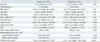
Dual-source computed tomography coronary angiography were successfully performed in all patients without side effects. The average HR and HR variability were 79 ± 9 bpm (range: 66-109 bpm) and 10.72 ± 4.88 bpm (range: 2-41 bpm) in group A, while the values for group B were 80 ± 11 bpm (range: 66-139 bpm) and 10.05 ± 5.69 bpm (range: 0-38 bpm), respectively. The mean BMI was 23.8 ± 2.7 (range: 19.2-29.8) in group A and 23.7 ± 2.5 (range: 19.1-29.2) in group B. The mean scan length was 12.08 ± 1.12 cm (range: 9.6-14.89 cm) in group A and 12.72 ± 1.88 cm (range: 9.8-14.5 cm) in group B. The acquisition time was 247 ± 25 ms (range: 194-417 ms) and 252 ± 22 ms (range: 215-307 ms) in groups A and B, respectively. There were no significant differences in age, BMI, HR, HRV, scan length, scan time or calcium score between the two groups (all p > 0.05). An overview of the results for groups A and B can be found in Table 1.
In total, there were 1842 coronary segments in group A and 1838 segments in group B. The inter-observer agreement for image quality rating was good (group A: kappa = 0.63; group B: kappa = 0.65). Image quality was rated as being excellent (score 1) in 92.3% (1701/1842) of segments in group A and 95.3% (1751/1838) of segments in group B; moderate in 6.1% (113/1842) of segments in group A and 3.0% (56/1838) of segments in group B; and non-diagnostic in 1.5% (28/1842) of segments in group A and 1.7% (31/1838) of segments in group B. There were no significant differences in image quality scores and proportion of non-diagnostic coronary artery segments between groups A and B (image quality scores: 1.064 ± 0.306 vs. 1.084 ± 0.327, respectively, p = 0.063; non-diagnostic coronary artery segments segment-based analysis 1.52% vs. 1.74%, respectively, p = 0.345; and patient-based analysis 7.5% vs. 6.7%, respectively, p = 0.812) (Table 2).
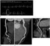
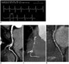
Ten patients had non-diagnostic images due to motion artifacts in group A. In five of these patients the motion artifacts occurred at the RCA and LCX because the acquisition phase fell on the R wave due to high HRV (Fig. 2). Examples of good image quality are displayed in Figure 1, while examples of typical non-diagnostic images are displayed in Figure 2. Table 2 summarizes the results of subjective image quality evaluation in both groups.
The average image noise was 21.4 ± 4.5 HU (range: 19-27 HU) and CNR was 12.1 ± 4.2 (range: 6.4-25.3) in group A, and the corresponding numbers were 20.9 ± 4.3 HU (range: 19-28 HU) and 13.8 ± 5.1 (range: 7.1-28.2) in group B. There were no significant differences in image noise or CNR between the two groups (Table 1).
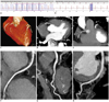
The average HRV was 2.29 ± 1.06 (95% CI 2.05-2.53) in patients of score 1, 5.17 ± 1.37 (95% CI 4.77-5.58) in patients of score 2, and 8.88 ± 1.53 (95% CI 7.71-10.07) in patients of score 3 in group A. In group B, the average HRV was 2.61 ± 1.85 (95% CI 2.23-2.99), 7.90 ± 3.97 (95% CI 6.47-9.34) and 11.22 ± 5.62 (95% CI 6.89-15.55) in patients of scores 1-3, respectively (Table 2). In group B, four patients' vessels were unassessable from motion artifacts induced by poor cooperation in breath-holding. Diagnostic evaluation of the entire coronary artery tree was obtained in qualified patients using flash mode in acquisition at 20% of the R-R interval (Fig. 3).
The DLP of DSCTCA examinations was 61.17 ± 7.24 mGy·cm in group A and 414.67 ± 61.71 mGy·cm in group B. The estimated radiation dose was 1.04 ± 0.16 mSv (range: 0.7-1.55 mSv) in group A and 7.05 ± 1.05 mSv (range: 5.68-11.92 mSv) in group B (p = 0.001) (Table 1).
DISCUSSION
There are three scan modes in high pitch DSCT examinations: spiral mode, sequential mode and flash mode (22). In spiral mode, the spiral DSCTCA scan technique delivers the X-ray throughout the entire cardiac cycle. Even with ECG-based tube current modulation, the radiation dose is still high and the effective radiation doses can be as high as 12 mSv (7). In sequential mode, the radiation exposure is turned on only with the optimal phase of the cardiac cycle and then turned off (23). This short time data acquisition can reduce the radiation dose substantially, resulting in an average effective radiation dose of 2.1-4.1 mSv (8, 24-26). Moreover, the "padding" technique allows for an additional exposure time before and after the optimal phase in the cardiac cycle. Overall, the high-pitch spiral mode provides a high diagnostic accuracy for the assessment of coronary stenosis with doses below 1 mSv (11).
The most challenging issue in CTCA examination is non-diagnostic images. The rate of non-diagnostic segments has been reported to be as high as 12% with 64-section CTCA (27), with a range of 1 to 6% with first generation DSCTCA (28-30) and around 10% of patients affected. The high-pitch spiral mode of DSCT permits data acquisition from the entire heart within one heart beat by continuous and fast table movement. Leschka et al. (11) found that 99% of evaluable coronary segments in CTCA performed with the flash mode were with HR of 60 bpm, while the initiation of the imaging was at 60% of the R-R interval. Sensitivity of 94% and specificity of 96% were reported for the detection of significant coronary artery stenosis. High HRs were thought to be problematic in these acquisitions, especially during the high-pitch mode needed for motion-free imaging in patients with a low HR. Thus, regular sinus rhythm and HR ≤ 60 bpm have been considered prerequisites for an adequate examination.
At higher HRs, motion artifacts will lead to a non-diagnostic image, predominantly affecting the mid RCA. The optimal time selection aims to minimize the coronary arterial motion artifacts, especially the artifacts in the mid to distal RCA. The optimal time may shift to systolic intervals in patients with high HRs as diastolic diastasis shortens and eventually disappears with increased HRs (31). With retrospectively ECG-gated DSCT, Adler et al. (32) found that the optimal systolic phase is between 35% and 50% of the R-R interval in patients with HR > 65 bpm. Araoz et al. (15) reported that optimal image sharpness of the coronary arteries was achieved at 35-40% of the R-R interval in patients with HR > 70 bpm. Recently, Goetti et al. (16) suggested initiation of the high-pitch CT scanning at 30% of the R-R interval in high-pitch DSCTCA in patients with high HRs (≥ 70 bpm) significantly improved the image quality of coronary arteries.
Using high-pitch DSCT, Goetti et al. (16) selected forty patients with HR ≥ 70 bpm and chose the 30% of of the R-R interval for triggering of imaging. They found that the systolic window for data acquisition from high-pitch DSCTCA in patients with high HRs (HR ≥ 70 bpm) significantly improves the quality of coronary artery imaging. Based on their finding, we performed CTCA with flash mode with the optimal phase setting at 20-30% of the R-R interval in patients with high HRs (> 65 bpm), and we demonstrated good diagnostic image quality of coronary arteries using these methods.
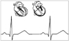
Mimimal motion artifacts occur in a phase of cardiac cycle with the least coronary artery motion. According to cardiac electrical mechanical coupling theory (33-36), the basic activity of the heart is electric and mechanical activity. After 40-60 ms of electric activity, the mechanical activity begins. Ventricle contraction includes three phases: isovolumetric contraction, rapid ejection and the slow ejection phase. The isovolumetric contraction phase lasts 50 ms, while the rapid ejection phase lasts around 100 ms. In the slow ejection phase, ventricle contraction is relatively slow and occurs about 150 ms after the QRS wave. For example, a high HR of 100 bpm (R-R interval: 600 ms), 30% of the R-R interval would be 180 ms. Data acquisition for high-pitch DSCTCA falls exactly in the slow ejection phase. Regarding the upper limit of HRs of the high pitch mode DSCTA, the scan time of high pitch mode CTCA is about 250 ms and the P-R interval is 120-200 ms. The isovolumetric contraction and rapid ejection phase lasts 150 ms. Thus, 150 + 250 + 200 ms = 600 ms (HR: 100 bpm). This gives an upper limit of HRs of 100 bpm with less coronary arterial motion artifacts via the high pitch mode DSCTCA. Figure 4 illustrates the most important features of the cardiac cycle and the best scan time of HP mode DSCTCA within a single heart beat in patients with high HRs.
A significant negative correlation between overall quality of images of the coronary arteries and HRV during scanning has been consistently reported since the introduction of MDCT cardiac imaging in clinical practice (37, 38). Using 64-Section CT, Leschka et al. (37) found HRV had a strong negative impact on image quality for all coronary segments and each coronary artery. Our data indicate that HRV is an important factor affecting the image quality of High-Pitch DSCTCA in patients with HRs, in agreement with previously reported work (37, 38). The average HRV in patients of score 3 were significantly higher than that of scores 1 and 2 in flash mode and spiral mode DSCTCA.
In our study, we found that four patients' vessels were unassessable using flash mode CTCA due to motion artifacts induced by poor cooperation in breath-holding. In contrast, the flash mode is still able to gain diagnostic evaluation of the entire coronary artery tree although patients cannot hold their breath. This suggests that the influence of respiration seems to be a minor concern for high-pitch spiral mode DSCTCA.
The reported effective doses for prospective CTCA were 2.1-4.3 mSv on 64-slice CT (39-41) and 1.5-2.5 mSv on the first generation DSCT (8, 29). Leschka et al. (11) reported a mean dose of 0.9 mSv in 35 patients that was investigated via high-pitch spiral CT, but with a less stringent control of tube voltage and current (320 mAs). In this study, the average effective radiation dose was about 1.04 mSv for the new flash spiral acquisition mode protocol. This dose is considerably lower than DSCT of 7.05 mSv for the retrospectively spiral scan in this study, and lower than retrospectively ECG-gated CTCA on DSCT reported by others (42). Moreover, it is substantially lower than that of first generation DSCT with reported values of 1.5-2.5 mSv on average (29). These results are similar to that of doses reported in other recent trials that evaluated high-pitch DSCT (9, 11, 43).
Study Limitations
There are several limitations to the study that we have performed. First, the image quality grading may have been inluenced by a subjectivity bias. Second, patients with irregular HRs were excluded, especially on atrial fibrillation or premature ventricular contraction. Third, in this study, we did not compare the results of the high-pitch acquisition protocol with sequential mode procols. Further studies will be required for a detailed analysis.
Conclusion
In conclusion, in patients with HRs > 65 bpm without cardiac arrhythmia, the prospectively high-pitch spiral acquisition mode with image acquired timing set at 20-30% of the R-R interval provides a similar image quality and low rate of non-diagnostic coronary segments to the retrospectively ECG-gated low-pitch spiral acquisition mode, with significant reduction of radiation exposure.

 XML Download