

 PDF
PDF ePub
ePub Citation
 Print
Print


INTRODUCTION
The benefits of endovascular treatment of posterior circulation aneurysms are well documented and it is a well established technique for the successful management of posterior inferior cerebellar artery (PICA) aneurysms. Surgical treatment of PICA aneurysms may be challenging due to their deep location and close relation with the lower brainstem and caudal cranial nerves (1-4). Infrequently, we encounter wide-necked, complex aneurysms of the PICA origin in our cerebrovascular practice and many of them still remain a significant therapeutic challenge in spite of improvements of endovascular techniques. Balloon- or stent-assisted coiling can be considered for selective occlusion of such wide-necked PICA aneurysms. In some PICA aneurysms, navigation of the stent through the ipsilateral vertebral artery (VA) may be very difficult or even impossible due to the unfavorable geometry of the aneurysm and/or parent vessel. Deconstructive techniques with PICA and/or VA occlusion have been considered the only possible endovascular option in these aneurysms (1).
This report describes the endovascular treatment and follow-up result of a wide-necked PICA aneurysm which was treated by retrograde stenting through the contralateral VA and vertebrobasilar junction (VBJ) with antegrade coil embolization preserving the VA and the PICA.
CASE REPORT
A 58-year-old man was referred to our hospital because of progressive gait disturbance and incontinence. The patient had undergone surgical clipping of a ruptured aneurysm at the right middle cerebral artery bifurcation in another institution 8 months earlier, and the case was complicated by a left-sided hemiparesis (grade IV) after surgery. At that time, an additional unruptured aneurysm at the left PICA origin had been diagnosed postoperatively by angiography. The PICA aneurysm had a wide neck and incorporated the PICA origin. The patient and his family were informed that the aneurysm posed a relatively high risk for rupture because the patient already had a subarachnoid hemorrhage from another aneurysm and the posterior circulation aneurysm could have a higher risk, because it would be very difficult to treat by either a surgical or endovascular approach.
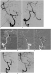
Hydrocephalus was diagnosed with computed tomography (CT) and treated with surgical placement of a ventriculoperitoneal shunt system in our hospital, and an uneventful follow-up period of 31 months. After the Enterprise stent system (Codman Neurovascular, Miami Lakes, FL, USA) became available, which is easier to deliver through the tortuous anatomy than other available stents, we planned a stent-assisted coil embolization of the PICA aneurysm. A follow-up cerebral angiography demonstrated a wide-necked aneurysm with a medially directed distal lobule (Fig. 1A, B). The aneurysm measured 6.1 mm in the long-axis diameter, 2.7 mm in the short-axis diameter, and 3.3 mm in height. There was no significant interval change in aneurysm size and shape. Because the left PICA coursed inferiorly, forming an acute angle with the proximal V4 segment of VA, it was decided that the Enterprise stent should be placed retrogradely with a contralateral (right) VA approach and coils should be placed into the aneurysm through the ipsilateral VA.
The patient was premedicated with 75 mg clopidogrel and 100 mg aspirin 72 hours before treatment. Under general anesthesia, we performed bilateral common femoral artery punctures. A Envoy guiding catheter (Codman Neurovascular, Miami Lakes, FL, USA) was introduced into the left VA and a 5 Fr Envoy into the right VA after administration of a 3000 U bolus of IV heparin. A "J-shaped", Prowler Select LP microcatheter (Codman Neurovascular, Miami Lakes, FL, USA) was then advanced through the 5 Fr Envoy guiding catheter into the right distal VA over a Synchro2 micro-guidewire (Boston Scientific, Natick, MA, USA) and navigated retrogradely into the left distal VA through the "acute-angled" VBJ with the help of a microcatheter tip shape (Fig. 1C). After the micro-guidewire was exchanged with exchangeable Agility-14 micro-guidewire (Codman Neurovascular, Miami Lakes, FL, USA), the microcatheter was retrieved and the Prowler Plus Select microcatheter (Codman Neurovascular, Miami Lakes, FL, USA) was advanced into the left distal PICA across the aneurysm neck for stent delivery (Fig. 1D).
Through this microcatheter, a 4.5 × 22 mm Enterprise stent was advanced into the left distal VA-PICA and placed over the aneurysm neck. Before stent deployment, a Prowler Select LP microcatheter was advanced through the ipsilateral VA and placed within the aneurysm. After partial coiling of the aneurysm dome with a 3 mm × 4 cm minicomplex Trulfill DCS Orbit detachable coil (Codman Neurovascular, Miami Lakes, FL, USA), the stent was deployed from the left proximal PICA to the distal VA (Fig. 1E). Subsequently, three more detachable coils (3 mm × 4 cm, 2.5 mm × 3 cm, and 2 mm × 2 cm minicomplex Trulfill DCS Orbit coils) were delivered into the aneurysm for further packing. A final control angiogram showed near-complete aneurysm occlusion and preservation of the PICA and VA flow (Fig. 1F). The patient recovered from anesthesia with an unchanged neurological status.
Clinical and angiographic follow-up at eleven months revealed complete occlusion of the PICA aneurysm (Fig. 1G) with no neurological change.
DISCUSSION
Surgical treatment of PICA aneurysms poses a great technical challenge and high morbidity/mortality like that of other posterior circulation aneurysms (1-4). First, PICA aneurysms are located deep and related closely to the lower cranial nerves. Secondly, the bony structures in the lateral foramen magnum region can be obstacles during surgery (2). Postoperative temporary or permanent cranial nerve palsies may occur in almost half of the cases with a prolonged need for intensive care and inherent risk of developing pneumonia (1, 5). Therefore, endovascular treatment has been considered the first treatment option for PICA aneurysms, especially for unruptured aneurysms if circumstances allow.
Endovascular treatment of wide-necked aneurysms of the PICA still remains a significant therapeutic challenge. In aneurysms incorporating the PICA origin, standard treatment techniques with preservation of the PICA flow may be impossible and require deconstructive techniques (1). However, because the deconstuctive technique should be considered the last resort, especially for an unruptured asymptomatic PICA aneurysm, application of the optimal modified technique may be necessary for selective occlusion of the aneurysm with preservation of the parent vessels. Since the advent of stent- or balloon-assisted techniques for treatment of wide necked intracranial aneurysms, several modified remodeling techniques have been introduced for treatment of wide necked aneurysms with a complex anatomy: the retrograde approach via communicating arteries using a balloon (6) or stent (7), combined use of stent and balloon ("balloon-in-stent technique") for fusiform aneurysms (8), "Y-stenting" (9, 10), stenting with contralateral vertebral approach via the vertebrobasilar junction (11), and other techniques. These innovative techniques have widened the indications for endovascular treatment of intracranial aneurysms.
The PICA has the most complex, tortuous, and variable course of the cerebellar arteries (4). In some cases, selective catheterization of the PICA for the placement of balloon or stent may be difficult or risky because of the acutely angled course of the PICA with respect to the VA. These difficulties and risks of stenting through the unfavorable vessel configuration were described by Kelly et al. (7) in their detailed observations. The stent delivery system often meets resistance in its passage through the acute angle beyond the aneurysm into the distal parent artery and may herniate into the aneurysm sac. This herniation may occur abruptly and can cause catastrophic procedural rupture. Accurate deployment of the stent across the aneurysm is also difficult because binding can occur within the stent delivery system. In our case, the catheterization for stenting using the ipsilateral approach was considered hazardous with a significant risk for aneurysm rupture due to the upward projection of the PICA aneurysm. Therefore, a retrograde approach into the PICA, instead of an ipsilateral approach, was chosen for stent placement to reduce rupture risk and to obtain complete neck coverage for secure coiling of the aneurysm.
Stent-assisted embolization of wide neck aneurysms in small and/or distal cerebral vessel is limited because periprocedural thrombus formation and parent artery occlusion may occur during or after the procedure. In a report regarding the successful use of Neuroform stent (Boston Scientific, Natick, MA, USA) placement into small cerebral vessels measuring < 2 mm in diameter (range, 1.1-1.8 mm), acute thrombosis occurred in 2 (25%) of the 8 patients, even though the thrombosis was resolved without symptomatic complications after abciximab administration (12). In our case, although we had great concern about acute or subacute thrombosis and delayed stenoocclusion of parent artery as the PICA was smaller (measuring 1.6 mm) than the minimal recommended parent vessel diameter of 2.5 mm for utilizing Enterprise stent, no symptoms occurred during the procedure and follow-up period.
The contralateral vertebral approach for stenting and coil embolization of the PICA aneurysm was first introduced in the literature by the Ecker et al (11). They successfully treated large, partially thrombosed aneurysms with a similar technique using a Neuroform stent after multiple unsuccessful attempts. In our opinion, their described aneurysm did not have as wide a neck as our case and could possibly have been treated with simple coiling or antegrade balloon- or stent-assisted coiling if a microcatheter had been navigated beyond the aneurysm.
Because of the acute angle of the VBJ in our case, we used the Enterprise stent system which had the ability to negotiate the tortuous anatomy (13). The high flexibility of the Enteprise stent system in combination of its delivery through a Prowler Plus Select microcatheter facilitated retrograde placement of the stent into the PICA. We believe that it would have been significantly more difficult to advance any other available stent into the PICA through the VBJ due to the acute angle.
Conclusion
Endovascular treatement of wide-necked aneurysms with a PICA origin can be very difficult or impossible when the proximal intracranial VA and PICA form an acute angle. In these cases, retrograde stenting through the VBJ into the PICA in conjunction with antegrade coiling of the aneurysm should be considered.
 XML Download