

 PDF
PDF ePub
ePub Citation
 Print
Print


INTRODUCTION
Non-compaction of the left ventricle is a rare congenital disorder thought to be caused by the arrest of compaction of the myocardial fibers during embryogenesis (1). The echocardiographic findings of this disorder have been reported in small series but CT and MR imaging findings have so far been less frequently reported (2-4). We describe the case of a patient with new-onset heart failure in whom cardiac CT (5) identified characteristic myocardial changes of the isolated left ventricular non-compaction and associated global and regional functional abnormalities, as well as atherosclerotic coronary artery disease.
CASE REPORT
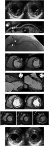
A 72-year-old man with a history of coronary artery disease and dilated cardiomyopathy was admitted to our hospital with abrupt onset of dyspnea for the last three days. Upon admission, atrial fibrillation was diagnosed on electrocardiogram (ECG). A chest CT showed mild pulmonary edema in both lungs and bilateral pleural effusion. An echocardiography performed on the day of admission, severe left ventricular systolic dysfunction was found and prominent trabeculation of the left ventricular cavity was described (Fig. 1A). The ejection fraction was decreased to 30%.
Cardiac CT was then performed to assess the progression of coronary artery disease previously noted on coronary catheter angiography (CAG) five years prior to the current admission and to further elucidate the ventricular abnormality suspected on echocardiography. Retrospectively ECG-gated cardiac CT was performed with a 64-slice MDCT system (Somatom Sensation 64 Cardiac, Siemens Healthcare, Forchheim, Germany). A standardized protocol with a collimation of 64 × 0.6 mm, gantry rotation time of 330 millisecond, and a pitch of 0.2 was used. Tube voltage was 120 kV with an effective tube current-time product of 770 mAseff and an ECG-dependent tube current modulation. The scan was contrast-enhanced with 65 ml of a non-ionic contrast medium (Ultravist; 300 mgI/ml, Bayer, Berlin, Germany), injected at 5 mL/s through an 18 G intravenous antecubital catheter, and using a tri-phasic contrast delivery protocol.
Cardiac CT showed 50% luminal stenosis in the first diagonal branch of the left anterior descending artery (Fig. 1B), which was in good correlation with the findings on the previous CAG (Fig. 1C). Functional cine reconstructions revealed thickening of the left ventricular wall with prominent trabeculations and deep intertrabecular recesses (Fig. 1D, E). Short axis images revealed that the intertrabecular recesses were filled with blood and were "milked" during systole (Fig. 1F). This was visually associated with hypokinesis of the lateral wall of the left ventricle (Fig. 1F). Global functional parameters were obtained on an automated image processing workstation (Aquarius, TeraRecon), which showed an ejection fraction of 34% in good correlation with the echocardiography results. To assess the underlying ischemic changes, contrast-enhanced cardiac MRI was performed on the next day, which failed to demonstrate delayed enhancement suggestive of chronic infarction but confirmed the extent and distribution of the left ventricular trabeculae and recesses, as well as the wall motion abnormalities seen on CT (Fig. 1G-I). The ejection fraction measured at MRI was 33%.
Since the clinical and imaging evaluation supported the diagnosis of heart failure associated with left ventricular non-compaction without significant coronary artery stenosis or ischemic changes, the patient received routine treatment for cardiac failure under which his ejection fraction subsequently improved to 40% on echocardiography (Fig. 1J).
DISCUSSION
Isolated non-compaction of the left ventricle is a rare congenital cardiomyopathy characterized by prominent trabeculae, deep intertrabecular recesses, and thickened myocardium with two distinct layers (compacted and non-compacted). It typically involves the left ventricle, although involvement of the right ventricle has also been reported. Patients may be asymptomatic or suffer from dyspnea, severe systolic dysfunction, arrhythmias, and embolic events secondary to atrial fibrillation (6). The prevalence of myocardial non-compaction among heart failure patients may be underestimated, probably as a result of the underutilization of imaging and lack of physician awareness (7) concerning this disorder, as in our case. This is in keeping with recent observations, which noted an increased rate of diagnosis of the left ventricular non-compaction with the use of cross-sectional imaging modalities (2, 7).
Traditionally, echocardiography has been used to establish a diagnosis of myocardial non-compaction, although CT and MRI provide better visualization of the trabeculations. Even so, the latter modalities have yet to be systematically evaluated in larger cohorts to assess their clinical utility (8). In our case, both CT and MRI clearly delineated the area over which the trabeculae were distributed and also confirmed the criteria for establishing a diagnosis of myocardial non-compaction (8, 9) (Fig. 1D-H). In addition, CT, which was initially performed for the evaluation of new-onset heart failure, enabled quantitative and qualitative assessment of global and regional ventricular function from the same dataset in addition to the morphological assessment for coronary artery disease and general cardiac morphology. Accordingly, the integrative nature of cardiac CT suggests an advantage of this modality over other imaging tests such as echocardiography and CMR (10) in this setting.
Although cardiac MRI is currently considered as the reference standard for morphologic and functional assessment of the heart owing to its noninvasive nature, reproducibility, and excellent temporal resolution, its use for coronary artery evaluation is limited.
In conclusion, in this patient, cardiac CT enabled the noninvasive diagnosis of left ventricular myocardial non-compaction, thus highlighting the potential utility of this imaging test for elucidating rare and unexpected etiologies of abrupt-onset heart failure as well as aiding in therapeutic planning. Further, this case illustrates the integrative nature of cardiac CT imaging, enabling the simultaneous evaluation of complex intracardiac pathology and ventricular function as well as for coronary artery morphology with a single test.
 XML Download