

 PDF
PDF ePub
ePub Citation
 Print
Print


INTRODUCTION
A chondromyxoid fibroma (CMF) is an uncommon benign cartilaginous tumor of bone that makes up less than 1% of all bone tumors and furthermore, is the least common of the cartilage tumors (1). This tumor classically occurs in the metaphyseal region of the long bones around the knees, but it also occurs in other long bones, short bones of the hands and feet, pelvis, ribs, scapula, and spinal column (2-5). This metaphyseal tumor in the tubular bones may extend into the adjacent epiphysis or into the diaphysis or both. A primary diaphyseal or epiphyseal origin for this tumor is rare (2-7). We present here a case of a CMF that occurred in the epiphysis of the proximal tibia with an open growth plate.
CASE REPORT
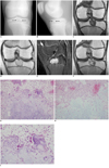
A 15-year-old boy presented with generally increasing pain in his left knee during over the previous month; he had no history of trauma. Upon physical examination, the boy complained of tenderness on the anterior aspect of his left knee. The results of routine laboratory studies were normal. The roentgenograms showed a 2.1×1.4×2.4-cm-sized, well defined, eccentric, osteolytic lesion with surrounding sclerosis in the proximal tibial epiphysis (Fig. 1A, B). In addition, there was no matrix calcification or periosteal reaction. The lesion slightly abutted the growth plate, which was still unfused. Upon magnetic resonance (MR) imaging, the lesion showed low signal intensity on the T1-weighted image, high signal intensity with a hypointense peripheral rim, and suspicious matrix calcifications on the proton-density and fat-suppressed T2-weighted images. Furthermore, there was no surrounding marrow edema. After intravenous administration of gadolinium diethylenetriamine pentaacetic acid (Gd-DTPA), the lesion enhanced heterogeneously with non-enhancing, hypointense foci (Fig. 1C-F). The preoperative presumptive radiologic diagnosis was chondroblastoma, and the differential diagnosis included enchondroma. A curettage and autologous bone graft were performed. The histologic examination of the excised tissue showed macrolobules with hypercellularity at the periphery of the lobules, and low cellular myxoid stroma at the center of the lobules (Fig. 1G-I). Chicken wire or granular calcifications were present, as well as woven bone formation. On the follow-up after three years, this tumor did not recur in clinical and radiological exams.
DISCUSSION
A CMF is a relatively rare, benign cartilaginous bone tumor that was first described in 1948 by Jaffe and Lichtenstein (8). It is most common in the second and third decades of life, with a reported age range from 4 to 87 years. The symptoms include slowly progressive pain, tenderness, swelling, and restriction of motion are observed (3, 5). Although CMF is typically located in a metaphyseal location, it may also extend to the epiphysis or diaphysis. A primary epiphyseal location is very rare; we were only able to identify four CMF cases occurred in the epiphysis of a long bone in the English language medical literature (4-7). Based on the roentgenograms, three of four cases were eccentric, osteolytic lesions with a sclerotic rim, whereas one of the tumors was expansile (4, 6, 7).
The MR imaging appearance of a CMF has been described in a few reports (9-11). On T2-weighted spin echo images, it has been described as having multilobular patterns and high signal intensity, and these features are similar to the characteristics found in hyaline cartilage bone tumors (9-11). However, for CMFs, the high signal intensity on the T2-weighted images is probably pathologically related to the abundant myxoid stroma maintained by thin fibrous tissue in a multilobular pattern; this tumor may show hyaline cartilage differentiation (4, 5, 9-11). The MR imaging appearance of CMF can be very heterogeneous; and, its polymorphologic appearance depends on the different degrees of cellularity and the presence of varying amounts of myxoid and fibrous tissue (10).
When CMF involves the epiphysis in adolescents, the lesion may be confused with chondroblastoma or enchondroma (6). Chondroblastoma usually originates in the epiphysis or apophysis of a long tubular bone and as a result, may traverse the growth plate and involve the metaphysis. The lesion is seen with low signal intensity on the T1-weighted spin echo images and variable (and often low) signal intensity on the T2-weighted images (12). Calcification in CMFs has been reported to occur in 2% and 16% cases seen on roentgenograms, but much less than those of chondroblastoma (30-50%) (2-4, 12). Enchondroma generally appear as lucent, well demarcated round or oval lesions with typical cartilaginous matrix mineralization, and occasionally, expansile with thinned cortical margins. In addition, the lesion shows characteristic high signal intensity foci of the hyaline cartilage on T2-weighted MR images. In very rare cases, the tumor occurs in an epiphysis where the lesion tends to involve almost the entire epiphysis in contrast to CMF (2, 6, 11). Giant cell tumor (GCT) is an epiphyseal lesion without calcification in a mature skeleton, and is seldom encountered in a patient younger than age 20 years or prior to epiphyseal closure (2). The tumor tends to invade and destroy the adjacent cortex to a greater degree as compared to a CMF.
As a benign lesion, a CMF is treated by curettage or excision. With curettage, a 13% to 25% recurrence rate has been reported. The recurrence rate has been reduced with the use of allograft bone or polymethylmethacrylate (3, 7). In the present case, there was no evidence of recurrence at three years post-operatively.
Histologically, a CMF consists of lobulated areas of myxomatous tissue separated by fibrocellular bands with little tendency for chondroid formation. Giant cells may be present in the fibrous regions. Many histological variations of this pattern occur, such that the lesion may be confused with other tumors or it may mimic a malignancy such as chondrosarcoma, fibromyxosarcoma or osteosarcoma (4, 5). Our case showed the characteristic histopathology of a CMF. Suspected calcification within the tumor was also observed on the MR images as well as microscopically. The plain radiographs failed to demonstrate calcification. The additional computed tomography (CT) was not performed to confirm the calcification. In our case, radiographic findings were consistent with the known imaging findings of epiphyseal CMFs and the MR imaging appearance was useful to distinguish the CMF from bone tumors of non-cartilage origin. However, an epiphyseal CMF is unusual and other bone tumors originating in the cartilage should be considered in the differential diagnoses.
In summary, we report a case of a CMF that occurred in the epiphysis of the proximal tibia. To our knowledge, this is the first report of MR imaging of an epiphyseal CMF in the English literature.
 XML Download