Abstract
Malignant mesothelioma (MM) is a relatively rare carcinoma of the mesothelial cells, and it is usually located in the pleural or peritoneal cavity. Here we report on a unique case of MM that developed in the chest, abdominal and pelvic walls in a 77-year-old female patient. CT and MRI revealed mesothelioma that manifested as a giant mass in the right flank and bilateral pelvic walls. The diagnosis was confirmed by the pathology and immunohistochemistry. Though rare, accurate investigation of the radiological features of a body wall MM may help make an exact diagnosis.
Malignant mesothelioma (MM) is a relatively rare but highly invasive tumor that arises from mesothelial cells of the pleura, peritoneum, pericardium or spermatic sheath. MM may present as the local or diffuse form. A typical MM usually presents as diffuse or multiple pleural or peritoneal nodules, but it rarely presents as a chest or abdominal wall mass. There have been only three cases of pleural or peritoneal MM presenting as a chest or abdominal wall mass reported in the medical literature (1-3), of which one case occurred in the thoracic wall as a local pleural MM, and the other two cases occurred in the abdominal wall. The giant MM reported in this current article simultaneously invaded the chest, abdominal and pelvic walls. To the best of our knowledge, this is the only reported case of MM that was simultaneously located in the chest, abdominal and pelvic walls, and the mass was larger than any previously reported case.
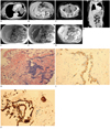
A 77-year-old female accidentally noticed a progressively enlarging lump in her right abdominal wall one year earlier and she didn't receive any treatment. Loose stools appeared for the past half year without any obvious weight loss. A month previously, the patient suffered from shortness of breath after physical activity and eventually she visited our hospital, where the physical examination revealed a giant irregular tumor in the right chest, abdominal and pelvic walls. The upper edge of the tumor reached the lateral margin of the right breast and the lower edge was at the level of the pubic symphysis. The mass was hard without tenderness and poorly circumscribed with limited mobility. The local skin temperature was normal. The patient was a housewife: she denied any history of exposure to asbestos dust and poisonous chemicals and she had no symptoms, including fever, sweating at night, cough and chest or abdominal pain. The routine blood tests and serum carcinoembryonic antigen (CEA), CA12-5 and CA19-9 were normal. The plain chest X-rays showed a large amount of pleural effusion in the right thorax accompanied with mediastinum shift. Ultrasound-guided drainage was performed after admission. Cytological examination of the pleural fluid revealed malignant tumor cells. Chest, abdominal and pelvic computed tomography (CT) showed a giant irregular soft-tissue mass in the right thoracoabdominal wall and bilateral pelvic walls that had extensively invaded the muscular layers of the wall and extended to the inside of the abdominal cavity with peritoneum thickening (Fig. 1A-D). The mass had poorly-defined margins. There was no clear demarcation between the mass and the surrounding body wall tissue. The abdominal and pelvic lesions extended to the subcutaneous tissue with spiculated margins. The tumor contained no calcium tissue and the tumor showed inhomogeneous slightly decreased attenuation with a CT value between 32 and 48 Hounsfield units on the unenhanced CT, and heterogeneous mild to moderate enhancement with a CT value that was increased 21-26 Hounsfield units on the contrast-enhanced CT. The pleural fluid causing the effusion was completely drained. There was no significant evidence of pleural thickening (Fig. 1A). No abnormal lesion was seen in both lungs. We noted ascites and part of the small bowel was dilated in the abdominal and pelvic cavity, but no soft-tissue tumor was seen inside (Fig. 1C, D). The mass on abdominal magnetic resonance imaging (MRI) was iso-intense on the precontrast T1-weighted images (Fig. 1E), heterogeneous iso- to hyper-intense on the T2-weighted images (Fig. 1F) and it was heterogeneously enhanced with multiple patchy non-enhancing necrotic areas after contrast medium administration (Fig. 1G). The abdominal wall part of the lesion was bigger and more inhomogeneous than the other parts. The tumor itself contained no fat, yet there were multiple patchy areas of fat tissue in the superficial part of abdominal lesion due to subcutaneous tissue infiltration, and surrounding subcutaneous tissue swelling was also noted on the fat-suppressed T2-weighted images.
Biopsy was performed and specimens were taken from three different sites of the abdominal wall mass. Hematoxylin & Eosin staining showed papillary and tubular malignant tumor cells and proliferating mesothelial cells that had extensively invaded the muscular tissue (Fig. 1H). The tumor cells were positive for high molecular weight cytokeratin (34βE12) (Fig. 1I), HBME-1, calretinin (Fig. 1J), Wilm's tumor-1 (WT-1) and vimentin, and the tumor cells were negative for thyroid transcription factor-1 (TTF-1) and villin. These results confirmed the diagnosis of MM.
As the tumor was very large and extensively invasive, surgical resection was not possible and radiotherapy was also not done. The patient was treated symptomatically and sadly, she died six months later due to gradual deterioration of her condition.
Malignant mesothelioma is a relatively rare tumor that arises from mesothelial cells. Pleural MM is the most common type of MM and the next most common type is malignant peritoneal mesothelioma. Most but not all MMs are related with occupational or environmental exposure to asbestos (4). The patient in our case denied any history of exposure to asbestos and poisonous chemicals. As the incidence of MM has tended to have gradually increased in the world, this malady has aroused more attention.
Malignant mesotheliomas presenting as a chest or abdominal wall mass are usually caused by direct invasion of the pleural or peritoneal MM. The present case first presented as a mass in the abdominal wall, and it had invaded the abdominal cavity. No significant evidence of pleural thickening was noted nor was any abnormal lesion seen in lungs. We concluded that the present case of MM was a peritoneal mesothelioma that directly invaded to the chest, abdominal and pelvic walls, and it became a giant mass.
According to the histological morphology, MM is divided into three sub-types: epithelial, sarcomatoid and mixed. The diagnosis of MM mainly depends on the pathology and immunohistochemistry. A body wall MM should be differentiated from various soft-tissue sarcoma, sarcomatoid and metastatic tumors. According to the literature (5), the main positive MM markers include HBME-1, 34βE12, calretinin, WT-1 and vimentin, and the negative markers mainly include TTF-1 and villin. The pathology and immunohistochemistry of the present case were consistent with the diagnosis of the mixed type MM, and the pathological feature of the muscular tissue being diffusely invaded corresponded with the CT and MRI findings.
In addition, imaging examinations also play an important role in the diagnosis, staging and follow-up treatment. A contrast-enhanced CT scan is the main imaging examination for MM (6). CT is able to completely scan the chest, abdomen and pelvis in a single process. This is helpful for determining the origin of MM, the scope of the pathologic invasion and making the differential diagnosis from various organ tumors. This advantage was also reflected in our case. MRI, which provides better soft-tissue contrast resolution than does other imaging modalities, allows accurate characterization of the mass and the relationship between the tumor and the surrounding structures, which may help distinguish between MM and other soft-tissue tumors of the body wall (7).
The imaging-based differential diagnosis of a body wall MM may mainly include other infiltrative lesions, such as abdominal fibromatosis, muscle lymphoma and metastatic tumors. Since this type of MM is rare and only a few cases have been reported with the CT images, there has been no detailed description of the body wall MM imaging features. According to the previous literature and the present case, the following radiological features may help make the radiological diagnosis and differential diagnosis of a body wall MM (7-10). First, the CT attenuation values of MM are somewhat heterogeneous and similar to or slightly lower than that of normal muscle on unenhanced CT, with a mild to moderate increase on contrast-enhanced CT. On T2-weighted images, MMs are compared with muscle with heterogeneous equal or slightly increased signal intensity. However, on the CT images, mostly abdominal fibromatosis and muscle lymphoma have relative homogeneous intermediate attenuation and moderate to marked enhancement; on T2-weighted images, the predominant signal intensity of MM lesions is intermediate, between that of muscle and that of subcutaneous fat, and the features that the T2-weighted images depict are hypo-intense bands that are not enhanced and these will help differentiate fibromatosis from MM. In addition, as the pathological examinations have shown, MMs have a few necrotic areas that are revealed on contrast-enhanced MRI, which will help differentiate body wall MM from metastatic tumors and infectious diseases. Second, a body wall MM is usually characterized by a poorly-defined margin that frequently extends and infiltrates along the body wall, and this type of MM is more aggressive than the other types of MM. Compared with them, most other soft-tissue masses and even metastatic tumors have a relatively clear margin and especially on the contrast-enhanced CT and MRI. Third, the body wall MM that probably arises from the pleura or peritoneum is often accompanied by pleural or peritoneal thickening and effusion.
In summary, we report here on a rare case of MM with a unique presentation. Although the diagnosis of MM depends on the pathology and the radiological examination findings are not specific, the features differentiating body wall MM from other soft-tissue infiltrative tumors are CT attenuation, MRI signal intensity, the pattern of contrast enhancement, diffuse involvement of the body wall and peritoneal or pleural thickening, which may help make an exact diagnosis.
Figures and Tables
Fig. 1
77-year-old female with malignant mesothelioma in chest, abdominal and pelvic walls.
A-D. Contrast-enhanced CT scan shows giant irregular soft-tissue mass in right thoracoabdominal wall and bilateral pelvic walls, and this has extensively invaded body wall, and mass extends to inside of abdominal cavity with peritoneal thickening. E-G. Mass is iso-intense on T1-weighted images (E), heterogeneous iso- to hyper-intense on T2-weighted images (F) and heterogeneously enhanced with multiple patchy non-enhancing necrotic areas on post-contrast fat-suppressed T1-weighted image images (G). H. Papillary malignant tumor cells and proliferous spindle cells diffusely invade into striated muscle (Hematoxylin & Eosin staining, × 100). I, J. Tumor cells are positive for 34βE12 (I) and calretinin (J) (immunohistochemistry, × 200).

References
1. Hayashi H, Notohara K, Yoshioka H, Matsuoka T, Ikeda H, Kagawa K, et al. Localized malignant pleural mesothelioma showing a thoracic mass and metastasizing to the stomach. Intern Med. 2010. 49:671–675.
2. Videtic GM. Primary malignant mesothelioma of the abdominal wall: complete response with radiotherapy alone. Technol Cancer Res Treat. 2008. 7:41–44.
3. Takeda T, Nishimura Y, Tsuchiya T, Nakata K, Takenaka K, Nakata H, et al. A large abdominal wall mass as an initial manifestation of malignant mesothelioma. Am J Med Sci. 2007. 333:218–220.
4. Bridda A, Padoan I, Mencarelli R, Frego M. Peritoneal mesothelioma: a review. MedGenMed. 2007. 9:32.
5. Kushitani K, Takeshima Y, Amatya VJ, Furonaka O, Sakatani A, Inai K. Differential diagnosis of sarcomatoid mesothelioma from true sarcoma and sarcomatoid carcinoma using immunohistochemistry. Pathol Int. 2008. 58:75–83.
6. Moore AJ, Parker RJ, Wiggins J. Malignant mesothelioma. Orphanet J Rare Dis. 2008. 3:34.
7. Dinauer PA, Brixey CJ, Moncur JT, Fanburg-Smith JC, Murphey MD. Pathologic and MR imaging features of benign fibrous soft-tissue tumors in adults. Radiographics. 2007. 27:173–187.
8. Chun CW, Jee WH, Park HJ, Kim YJ, Park JM, Lee SH, et al. MRI features of skeletal muscle lymphoma. AJR Am J Roentgenol. 2010. 195:1355–1360.
9. Kawashima A, Goldman SM, Fishman EK, Kuhlman JE, Onitsuka H, Fukuya T, et al. CT of intraabdominal desmoid tumors: is the tumor different in patients with Gardner's disease? AJR Am J Roentgenol. 1994. 162:339–342.
10. Kim HJ, Lee HK, Seo JJ, Shin JH, Jeong AK, Lee JH, et al. MR imaging of solitary fibrous tumors in the head and neck. Korean J Radiol. 2005. 6:136–142.


 PDF
PDF ePub
ePub Citation
 Print
Print


 XML Download